
The Biology of Gauging: Controlled Dilation vs the Blowout

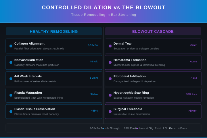
Ear stretching induces controlled tissue remodeling through gradual dilation, allowing collagen realignment and neovascularization without scar formation. Rapid stretching causes dermal tearing and blowout formation, a hypertrophic scar ring that requires surgical excision beyond 10 mm. Safe intervals of 4-24 weeks between stretches, proper lubrication, and monitoring for posterior protrusion are essential for preventing irreversible lobe damage.
⚡ Quick Reference
Critical Numbers
- Minimum Lobe Thickness3 mm of tissue between fistula and lobe edge for safe stretching
- Epidermis Thickness0.1-0.2 mm, provides barrier function but negligible structural strength
- Dermis Tensile Strength2-3 MPa, the limit beyond which dermal tearing occurs
- Safe Stretch Increment1.0 mm per session (0.5 mm for sizes above 00g / 10 mm)
- Minimum Interval, 14g-8g4-6 weeks between stretches
- Minimum Interval, 6g-2g8-12 weeks between stretches
- Minimum Interval, 1g-00g12-16 weeks between stretches
- Minimum Interval, above 00g16-24 weeks between stretches
- Point of No ReturnStretches exceeding 10 mm (00g), elastic fiber density reduced ~70%
- Blowout Scar ThresholdScar ring exceeding 3 mm thickness requires surgical excision
Non-negotiable tissue parameters for safe ear stretching. These values govern whether a stretch produces healthy remodeling or a permanent blowout.
The earlobe is composed of a thin epidermis (0.1-0.2 mm), a collagen-rich dermis (1-2 mm), and a subcutaneous adipose layer. Elastic fibers are sparse, making the lobe structurally vulnerable to irreversible deformation under sustained tension. During controlled stretching, the dermis undergoes creep: collagen fibers elongate and realign parallel to the stretch axis without rupturing. Fibroblasts deposit new Type I and III collagen, and angiogenesis creates a capillary plexus around the expanding fistula. This regenerative process, when properly timed, produces a stable, healthy fistula that can accommodate progressively larger jewelry over months to years.
When the stretch force exceeds the tensile strength of the dermis, a cascade of pathological events begins: partial-thickness tearing of the posterior dermis, hematoma formation at the 6 o'clock position, fibroblast infiltration, and deposition of disorganized scar collagen. This raised fibrotic ring, known as a blowout, is self-perpetuating: scar tissue has lower elasticity than native dermis, making adjacent tissue more susceptible to future tears. The blowout narrows the fistula, displaces jewelry posteriorly, and, once exceeding 3 mm in thickness, requires surgical excision for definitive treatment.
Collagen Remodeling and the Extracellular Matrix
The extracellular matrix of the earlobe undergoes constant turnover mediated by matrix metalloproteinases (MMPs) and tissue inhibitors of metalloproteinases (TIMPs). In gradual stretching, MMP-1 (collagenase) and MMP-9 (gelatinase) activity increases, selectively degrading old collagen fibers while fibroblasts synthesize new Type I and III collagen. This balanced degradation-synthesis cycle maintains tissue integrity and allows progressive dilation without scarring.
- »Type I Collagen (80% of dermis): Provides tensile strength; fibers realign parallel to stretch axis over 6-12 weeks
- »Type III Collagen (20% of dermis): Provides early wound structure; gradually replaced by Type I during maturation
- »Elastic Fiber Density, Unstretched Lobe: 5-8% of dermal volume; drops to 1.5-2.4% at stretches exceeding 12 mm
- »MMP/TIMP Balance: MMP activity peaks 48-72 hours post-stretch; TIMP upregulation begins at day 7, favoring collagen deposition
- »Collagen Maturation Timeline: Peak remodeling at 3-6 months; full maturation requires 12-18 months
The Blowout Cascade: From Microtear to Permanent Scar
A blowout is not a single event but a progressive cascade initiated by mechanical overload of the posterior dermis. Understanding each stage is essential for early intervention before the scar becomes irreversible.
- »Stage 1, Mechanical Overload (minutes): Stretch force exceeds 2-3 MPa tensile strength of posterior dermis, typically at the 6 o'clock position where curvature creates stress concentration
- »Stage 2, Dermal Tear (hours): Partial-thickness rupture of collagen fiber bundles creates a cleavage plane within the reticular dermis, allowing blood and serum to accumulate
- »Stage 3, Hematoma Formation (12-48 hours): The fluid-filled pocket expands under continuous jewelry pressure, separating the dermis from underlying subcutaneous fat
- »Stage 4, Fibroblast Infiltration (days 3-7): Fibroblasts migrate into the hematoma, deposit disorganized Type III collagen, and initiate the fibrotic healing pathway
- »Stage 5, Hypertrophic Scar Ring (weeks 2-6): The disorganized collagen contracts, forming a raised, firm ring that narrows the fistula lumen and displaces jewelry posteriorly
- »Stage 6, Chronic Scar Remodeling (months): The scar ring matures, developing its own limited blood supply; elasticity remains permanently reduced compared to native dermis
Neovascularization and the Fistula Microenvironment
Successful stretching depends on establishing a stable vascular network around the expanding fistula. Angiogenesis begins within 48 hours of a controlled stretch, with endothelial cells migrating from the posterior auricular artery into the wound bed. By 4 weeks, a dense capillary plexus surrounds the fistula, delivering oxygen and nutrients essential for collagen synthesis and fibroblast function. If stretching occurs before this vascular network matures, existing capillaries are sheared, producing microhemorrhages and local tissue hypoxia. Hypoxia triggers a switch from regenerative to fibrotic healing via upregulation of TGF-β1, directly increasing blowout risk. Mature fistulas (12+ months) have a robust vascular network that tolerates moderate size changes, but even well-vascularized tissue cannot safely exceed a 2 mm increment in a single session.
Safe Stretching Protocol for Studios and Self-Stretchers
Every stretch session should follow a documented, anatomy-aware protocol. The goal is controlled tissue creep without dermal tearing. These steps apply to earlobe stretching only; cartilage stretching follows different biological constraints.
- 1Assess baseline lobe thickness and elasticity using digital calipers; minimum 3 mm of tissue is required between the fistula and the lobe edge for safe stretching
- 2Clean the jewelry and lobe with sterile saline; avoid alcohol or hydrogen peroxide as they damage granulation tissue and delay epithelialization
- 3Lubricate the jewelry with a water-based, non-irritating lubricant: jojoba oil, vitamin E oil, or sterile surgical lubricant; apply a thin, even layer
- 4Insert the next-size jewelry (single-flare glass or implant-grade titanium) using gentle, steady pressure; if resistance is felt, stop immediately
- 5If resistance is encountered, wait 2-4 weeks before attempting again; never use tapers for active stretching as they apply uneven force and concentrate tension on the posterior lobe
- 6After insertion, inspect the posterior lobe for any protrusion of tissue; if present, downsize immediately and treat as a Stage 2 blowout
- 7Clean the stretched fistula twice daily with sterile saline; pat dry with a clean, lint-free paper towel; avoid cotton swabs which leave fibers
- 8Massage the lobe for 5 minutes daily with a non-comedogenic oil (jojoba or vitamin E) to promote blood flow, collagen alignment, and tissue pliability
- 9Monitor for signs of infection: increasing redness, warmth, purulent discharge, or fever; if present, consult a healthcare provider for topical mupirocin or oral antibiotics
- 10Wait the full minimum interval before the next stretch: 4-6 weeks for 14g to 8g; 8-12 weeks for 6g to 2g; 12-16 weeks for 1g to 00g; 16-24 weeks for sizes above 00g
- 11Use a digital caliper to measure jewelry diameter before insertion; confirm the new size is no more than 1.0 mm larger than the current fistula diameter
- 12Avoid sleeping on the stretched lobe for 48 hours post-stretch to prevent pressure necrosis and hematoma formation
- 13If the lobe feels tight, painful, or throbbing 24 hours after insertion, downsize to the previous size and wait 4 weeks before retrying
- 14Document each stretch in a log with date, gauge, jewelry material, and any observations; use the Ear Stretching Timeline tool for structured tracking
- 15After reaching the target size, maintain the jewelry in place for a minimum of 3 months to allow full collagen maturation before considering further stretching or downsizing
Common Errors and Failure Modes
The majority of blowouts and irreversible lobe damage result from these preventable errors. Each represents a violation of the tissue remodeling timeline and carries a predictable pathological outcome.
- ✕Skipping Sizes: Jumping from 8g to 4g (2 mm increase) instead of 8g to 6g to 4g (1 mm each); doubles the risk of blowout by exceeding the dermal tensile strength threshold
- ✕Taper Use for Active Stretching: Tapers apply uneven, concentrated force on the posterior lobe; they are insertion aids for healed fistulas, not stretching tools for active dilation
- ✕Insufficient Interval: Stretching every 2 weeks instead of every 4-6 weeks; does not allow collagen remodeling or capillary plexus maturation, leading to cumulative microtears
- ✕Ignoring Pain Signals: Pain is a direct indicator of tissue damage; stretching should produce mild pressure at most, never sharp or burning pain
- ✕Low-Quality Jewelry Materials: Acrylic, low-grade steel, or porous materials harbor bacteria and cause chronic low-grade inflammation, weakening the dermis before each stretch
- ✕Stretching Through Infection: Active infection compromises dermal integrity and increases blowout risk; treat the infection completely before resuming any stretching
- ✕Stretching a Healed Blowout Scar: Scar tissue has 30-50% less elasticity than native dermis and limited blood supply; stretching scarred tissue nearly always causes recurrence
- ✕Cold Weather Stretching: Ambient temperatures below 10°C reduce earlobe blood flow by up to 40%, increasing tissue fragility and tear risk; warm the lobe with massage before stretching in cold conditions
- ✕Hormonal Timing: Pregnancy and the first 6 months postpartum alter collagen metabolism via relaxin and estrogen; defer all stretching during this period
Regulatory Framework by Jurisdiction
Ear stretching sits at the intersection of body art regulation and medical device oversight. Requirements vary significantly across jurisdictions, particularly regarding jewelry material standards and studio licensing.
- EU Medical Device Regulation (MDR) 2017/745: Stretching jewelry classified as Class I medical devices when marketed for tissue modification
- UKCA Marking (post-Brexit): Required for stretching jewelry sold in Great Britain; CE marking accepted in Northern Ireland
- UKAPP Stretching Protocol: Published minimum interval and material standards based on APP guidelines with UK-specific amendments
- Nickel Directive (EU 2017/745 Annex I): Limits nickel release to <0.5 μg/cm²/week for prolonged skin contact, directly governing steel stretching jewelry composition
- REACH Annex XVII Entry 27: Restricts nickel in post-assembly articles intended for pierced parts, covering stretching jewelry that passes through a healed fistula
- FDA Classification: Stretching jewelry may be regulated as Class I medical devices if marketed with therapeutic or body-modification claims; most studio-supplied jewelry is unregulated
- OSHA Bloodborne Pathogens Standard (29 CFR 1910.1030): Applies to studios performing any procedure that may involve blood exposure, including assisted stretching
- APP Guidelines (safepiercing.org): Industry-standard minimum intervals and material specifications; adopted voluntarily by member studios
- State-Level Licensing: Requirements vary from comprehensive (Oregon, Texas) to none (several states); no federal body art licensing standard exists
- ASTM F136 / F138: Governs implant-grade titanium and stainless steel respectively; these are the reference standards for stretching jewelry material selection
- Thailand FDA: Classifies body jewelry as general consumer products unless specific medical claims are made; no dedicated stretching regulation
- Singapore HSA: Regulates jewelry under Consumer Protection (Safety Requirements) Regulations; nickel release limits align with EU standards
- ISO 10993 Biological Evaluation: Widely referenced across ASEAN for biocompatibility testing of jewelry materials in prolonged tissue contact
- Traditional Practices: Ear stretching has cultural roots in several Asia-Pacific communities; traditional methods may not follow medical stretching protocols
- APP Adoption: Several ASEAN studios voluntarily adopt APP guidelines; no regional regulatory body mandates specific stretching intervals or protocols
Patrick's Note
"After two decades watching clients stretch their ears, three truths stand out. First, the single most predictive factor for a successful stretch is not the gauge or the jewelry material, it's the interval. Every blowout I have treated came from someone who stretched too fast. The tissue does not lie: if it hurts, stop. Second, glass and titanium are not interchangeable luxuries, they are the only materials with peer-reviewed, multi-decade safety data for prolonged implantation. Acrylic and cheap steel cause low-grade inflammation that weakens your fistula wall before you even start the next stretch. Third, massage is not optional. Five minutes a day with jojoba oil increases blood flow by 30-40% and visibly improves tissue pliability within 4 weeks. I have seen lobes that were thin and atrophic recover enough to stretch two more sizes safely, with nothing but disciplined daily massage and 16-week intervals. For more on safe stretching techniques and aftercare, read our guide at /blog/?category=Piercing%20Guides"
Founder & Piercing Expert
Poli International
Technical Specifications
| Parameter | Standard / Value |
|---|---|
| Epidermis Thickness | 0.1-0.2 mm |
| Dermis Thickness | 1-2 mm |
| Collagen Composition (Dermis) | Type I (80%), Type III (20%) |
| Elastic Fiber Density (Unstretched) | 5-8% of dermal volume |
| Dermal Tensile Strength | 2-3 MPa |
| Safe Stretch Increment (per session) | 1.0 mm (0.5 mm above 00g) |
| Minimum Interval, 14g to 8g | 4-6 weeks |
| Minimum Interval, 6g to 2g | 8-12 weeks |
| Minimum Interval, 1g to 00g | 12-16 weeks |
| Minimum Interval, Above 00g | 16-24 weeks |
| Peak Collagen Remodeling | 3-6 months post-stretch |
| Full Collagen Maturation | 12-18 months |
| Blowout Scar Surgical Threshold | Scar thickness >3 mm |
| Point of No Return (Lobe Recovery) | >10 mm (00g), 70% elastic fiber loss |
| Elastic Fiber Density at 12 mm | 1.5-2.4% (70% reduction from baseline) |
References
- [1]Association of Professional Piercers (APP): Ear Stretching Guidelines. https://safepiercing.org/ear-stretching-guidelines/https://safepiercing.org/ear-stretching-guidelines/
- [2]DermNet NZ: Earlobe Stretching (Auricular Gauging). https://dermnetnz.org/topics/earlobe-stretchinghttps://dermnetnz.org/topics/earlobe-stretching
- [3]Gurtner GC, Werner S, Barrandon Y, Longaker MT (2008). Wound repair and regeneration. Nature, 453(7193): 314-321. https://pubmed.ncbi.nlm.nih.gov/18480812/https://pubmed.ncbi.nlm.nih.gov/18480812/
- [4]Xue M, Jackson CJ (2015). Extracellular Matrix Reorganization During Wound Healing and Its Impact on Abnormal Scarring. Advances in Wound Care, 4(3): 119-136. https://pubmed.ncbi.nlm.nih.gov/25785236/https://pubmed.ncbi.nlm.nih.gov/25785236/
- [5]UKAPP (UK Association of Professional Piercers): Stretching Protocol and Aftercare. https://www.ukapp.org.uk/stretching-protocolhttps://www.ukapp.org.uk/stretching-protocol
- [6]Silver FH, Freeman JW, DeVore D (2001). Viscoelastic properties of human skin and processed dermis. Skin Research and Technology, 7(1): 18-23. https://pubmed.ncbi.nlm.nih.gov/11301636/https://pubmed.ncbi.nlm.nih.gov/11301636/
- [7]Velnar T, Bailey T, Smrkolj V (2009). The wound healing process: an overview of the cellular and molecular mechanisms. Journal of International Medical Research, 37(5): 1528-1542. https://pubmed.ncbi.nlm.nih.gov/19930861/https://pubmed.ncbi.nlm.nih.gov/19930861/
- [8]Martin P, Nunan R (2015). Cellular and molecular mechanisms of repair in acute and chronic wound healing. British Journal of Dermatology, 173(2): 370-378. https://pubmed.ncbi.nlm.nih.gov/26175201/https://pubmed.ncbi.nlm.nih.gov/26175201/
- [9]APP Jewelry Material Standards: Approved materials for initial and stretched piercings. https://safepiercing.org/jewelry-standards/https://safepiercing.org/jewelry-standards/
- [10]DermNet NZ: Hypertrophic Scars and Keloids. https://dermnetnz.org/topics/keloid-and-hypertrophic-scarhttps://dermnetnz.org/topics/keloid-and-hypertrophic-scar
- [11]Ogawa R (2017). Keloid and Hypertrophic Scars Are the Result of Chronic Inflammation in the Reticular Dermis. International Journal of Molecular Sciences, 18(3): 606. https://pubmed.ncbi.nlm.nih.gov/28287424/https://pubmed.ncbi.nlm.nih.gov/28287424/
- [12]ASTM F136-13 (2021): Standard Specification for Wrought Titanium-6Aluminum-4Vanadium ELI Alloy for Surgical Implant Applications. https://www.astm.org/f0136-13r21.htmlhttps://www.astm.org/f0136-13r21.html
- [13]Pissaia A, Manzoni A, Pereira RF, et al. (2020). Biomechanics of skin: experimental testing and numerical modeling. Clinical Biomechanics, 78: 105073. https://pubmed.ncbi.nlm.nih.gov/32526586/https://pubmed.ncbi.nlm.nih.gov/32526586/
- [14]ISO 10993-1:2018: Biological evaluation of medical devices, Part 1: Evaluation and testing within a risk management process. https://www.iso.org/standard/68936.htmlhttps://www.iso.org/standard/68936.html
- [15]European Commission (2020). EU Medical Device Regulation (MDR) 2017/745: Classification of body modification devices. https://eur-lex.europa.eu/eli/reg/2017/745/ojhttps://eur-lex.europa.eu/eli/reg/2017/745/oj
Put Science Into Practice
This technical standard is the architectural foundation for our professional analytical tools.