Shop
Clinical Physics

Are Body Piercings Magnetic or Conductive? MRI Safety Facts

PP

Chief Engineer

Patrick Poli

Journal Date

2026-08-09

Last Reviewed

2026-10-11

Executive Summary. An MRI scanner is the strongest magnetic environment most people will ever enter. At 1.5 and 3 tesla, the static field exerts translational forces and torques on any ferromagnetic object, radiofrequency pulses can induce current in conductive jewelry, and even non-magnetic metals distort the image through susceptibility effects. The clinical physics explains why radiology departments insist on jewelry removal and which materials are genuinely safer.

The Static Field: Force and Torque

The static field B0 of an MRI scanner is measured in tesla. One tesla is roughly 20,000 times the Earth's surface field, and clinical scanners run at 1.5 T or 3 T, with research systems reaching 7 T. Two mechanical effects act on jewelry: translational force and torque.

Translational force pulls ferromagnetic objects toward the strongest field region, which in a superconducting magnet is the bore center. This is the projectile hazard. A loose steel barbell or ring can accelerate across the room or across the patient. The force scales with the field gradient, and the gradient is steepest at the bore entrance, which is why the danger zone starts before the patient is fully inside.

Torque rotates a magnetized object to align with the field. A ferromagnetic curved barbell or captive ring will try to twist in place. For a healed piercing this is uncomfortable but survivable; for a fresh fistula, the twisting can tear tissue and disrupt healing. Dedini et al. demonstrated at 1.5, 3, and 7 T that attraction forces scale dramatically with field strength, which is why behavior that is acceptable at low field can be dangerous at 3 T.
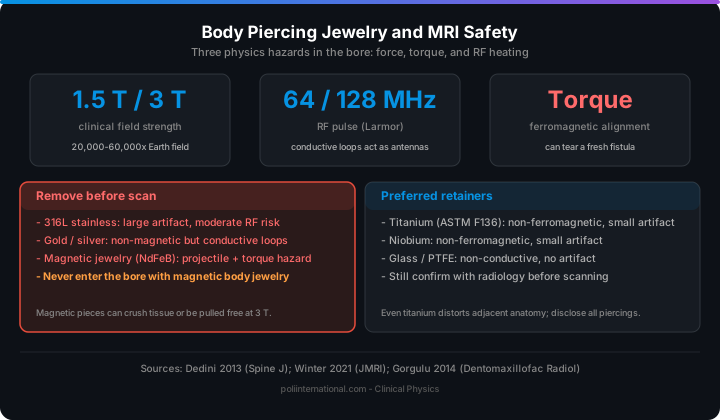
MRI safety of body piercing jewelry: ferromagnetic torque, RF heating, and susceptibility artifacts by material
Infographic: which piercing jewelry materials are safe inside an MRI scanner.

RF Heating: The Jewelry-as-Antenna Problem

MRI uses radiofrequency pulses at the Larmor frequency, roughly 64 MHz at 1.5 T and 128 MHz at 3 T. Conductive structures inside the bore can couple to these pulses and act as antennas. When a conductor forms a closed loop, a circular barbell, a ring, or a clicker, the RF field induces circulating current. Current density concentrates at small cross-sections such as the bead seat or the closure, and resistive losses appear as heat.

Winter et al. reviewed MRI-related heating of implants and devices and found that heating depends on geometry and position more than on material conductivity alone. The dominant variable is how well the conductor couples to the RF field, which is why a thin titanium ring can heat more than a thick steel bar if the ring forms a resonant loop. Görgülü et al. measured meaningful heating of small metallic orthodontic brackets at 3 T, and piercing jewelry is the same class of object, a small conductive loop at the body surface. Most scans are short enough that the temperature rise stays subclinical, but a localized burn at the piercing site is a documented risk, especially for jewelry that forms a complete conductive loop.

Susceptibility Artifacts: Why Non-Magnetic Is Not Invisible

Even non-ferromagnetic metals distort the magnetic field because their magnetic susceptibility differs from tissue. The local inhomogeneity dephases nearby protons and produces a signal void, the familiar black crescent around metal in an MRI image. Titanium has low susceptibility and produces a modest artifact. Stainless steel produces a larger one. Gold and silver, though non-magnetic, are highly conductive and distort the local field enough to matter.

The artifact matters even when the jewelry sits outside the primary region of interest. A tragus piercing during brain imaging or a navel barbell during lumbar spine imaging can degrade diagnostic quality enough to force a repeat scan. Li et al. reported tragus piercing as a risk factor during MRI precisely because of these interactions.

Material-by-Material Risk Assessment

MaterialFerromagnetic?RF loop riskArtifactMRI guidance
316L stainless steelMostly no (austenitic)ModerateLargeRemove
Titanium (ASTM F136)NoLowSmallPreferred retainer
NiobiumNoLowSmallPreferred retainer
Gold / silverNoModerateModerateRemove
Magnetic jewelry (NdFeB)Strongly yesHighSevereNever enter bore
Glass / PTFE retainersNoNoneNoneAcceptable

Clinical Takeaways

1. Remove all piercing jewelry before an MRI unless the imaging region is remote from the jewelry and the piece is verified non-ferromagnetic.
2. For piercings that cannot be removed, such as healing or tight-fitting jewelry, use glass or PTFE retainers, which are non-conductive and artifact-free.
3. Never scan with magnetic jewelry. Neodymium magnets are strongly ferromagnetic and experience both projectile force and torque at 3 T.
4. Titanium and niobium are the best metal choices for MRI-adjacent wear, but they still produce artifacts in nearby anatomy.
5. Tell the technologist about every piercing, including nipple and genital jewelry, because these are the ones most often missed in screening.
6. A healed piercing can close within hours without jewelry, so plan removal with a taper or retainer instead of gambling on the scan.


FAQ


Can I wear titanium piercing jewelry in an MRI?


Titanium is non-ferromagnetic and is generally considered MRI-safe for scanning, but it is not artifact-free. Commercially pure titanium and Ti-6Al-4V alloy have very low magnetic susceptibility, so the translational force and torque are negligible; however, the material will still produce a localized signal void due to susceptibility differences with tissue, which can degrade image quality if the piercing lies within the region of interest. A thin titanium ring that forms a closed conductive loop still carries an RF heating risk, as geometry and loop coupling are the dominant variables for heating, not material conductivity alone.


What happens if I forget to remove my metal piercing before an MRI?


Depending on the material, you risk projectile injury from the static field's translational force, torque-induced tissue tearing, or localized RF burns from induced currents. At 1.5 T, the force on a ferromagnetic steel barbell can exceed its weight several times over, and at 3 T that force scales up dramatically as demonstrated by Dedini et al., which is why a loose piece of steel can accelerate toward the bore center and become a projectile. Non-ferromagnetic but conductive metals like gold, silver, or copper rings can still couple to the RF field at 64 MHz (1.5 T) or 128 MHz (3 T) and heat enough to cause a burn, so radiology staff will typically halt the scan to have the jewelry removed.


Is 14k gold piercing jewelry safe to wear during an MRI?


Gold is not ferromagnetic, so it will not be pulled toward the magnet, but it is highly conductive and creates meaningful susceptibility artifacts that can obscure the image. More importantly, a gold ring or clicker forms a closed conductive loop that can act as an antenna for RF pulses; Görgülü et al. measured significant heating in small metallic structures at 3 T, and gold's low resistance can lead to higher circulating current and localized thermal injury. Even a simple gold stud without a closed loop can heat at the contact point with skin, so gold jewelry should be removed before a scan.


Will a surgical steel belly button ring set off the metal detector at an MRI facility?


Possibly, but the bigger concern is that many "surgical steel" or "316L stainless steel" pieces are not purely austenitic and may contain ferromagnetic inclusions from manufacturing or cold working. That residual ferromagnetism is enough for the magnet's translational force to rotate the curved barbell or pull it forward, torquing the fistula and potentially tearing fresh tissue; the attraction scales with field strength, so a piece that feels safe at 1.5 T can become dangerous at 3 T. Because the force depends on the exact alloy composition and manufacturing process, radiology departments cannot certify it as safe without testing, which is why the rule is "when in doubt, remove it."


Why does my MRI report mention an artifact from my nose piercing even though it was outside the scanning area?


Susceptibility artifacts are not confined to the immediate location of the metal; the local field inhomogeneity extends beyond the jewelry and dephases protons in surrounding tissue, creating a signal void that can appear as a black crescent over a wide field of view. Even a tragus piercing, as reported by Li et al., can degrade brain imaging quality, and a navel barbell can produce artifacts during lumbar spine scans because the distortion scale depends on the magnetic susceptibility difference, which for silver, gold, or stainless steel is large enough to affect adjacent slices. At 3 T, the artifact size increases with field strength, so a piercing that is "small" in size can still force a repeat scan if it overlaps the region of diagnostic interest.


6. MRI Safety by Material: Is Your Piercing Magnetic or Conductive?

People ask me two questions more than any others: are body piercings magnetic, and are piercings conductive? The honest answer depends entirely on the alloy. "Acier inoxydable", stainless steel, is a blanket term, not a specification. Most 316L surgical steel is austenitic and effectively non-ferromagnetic, which is why you can walk a basic steel ring past a fridge magnet without drama. But cheap jewellery and some "surgical steel" imports contain ferritic or martensitic phases, and those respond to a static field. That distinction matters, and magnetic response is only one of three hazards in the scanner.

Conductivity is the second. Even a metal that is not pulled by the magnet can still heat. At 1.5 and 3 T, radiofrequency pulses induce current in any conductive loop, a captive bead ring, a barbell with a snug ball, a nose hoop. The tighter the loop and the closer it sits to the coil, the higher the local specific absorption rate. This is why "non-magnetic" and "MRI-safe" are not synonyms, and why I never let a supplier conflate them. Titanium is non-ferromagnetic and comparatively low-risk for heating, but it still produces susceptibility artifacts that smear the image around the piercing site. Gold behaves similarly, and 14k or 18k alloys can contain enough ferromagnetic trace content to twist under torque.

My own position, after 25 years making this product: if you are heading into an MRI and cannot remove the jewellery, the retainer material is the decision that actually matters. BioFlex® is PP-R, a medical-grade plastic, not an elastomer, not PTFE, not silicone. It is non-metallic, so there is no ferromagnetic torque and no RF-induced current path. It does not generate the susceptibility artifacts metal does. That is the whole reason I developed it, and it is why the same material people wear for years in mouth, eyebrow, and cartilage piercings is also the sensible choice for imaging day.

Two practical rules before you scan. First, tell the radiographer exactly what you are wearing, where, and in what material, do not let them guess. Second, if the piercings are within the scan field, replace them with PP-R retainers and confirm retention before you enter the room, because a loose retainer is its own problem. For a material-by-material comparison before your appointment, work through our MRI retainer selection guide and check the imaging considerations for each option. And if you want the full materials breakdown behind these claims, start with BioFlex®.


Mind map · Are Body Piercings Magnetic or Conductive? MRI Safety Facts

  • Body Piercing MRI Safety
    • Metal Jewelry Hazards

      Magnetic resonance procedures subject metal body jewelry to magnetic displacement forces and radiofrequency-induced heating inside the scanner.

      • Mechanical Forces
        • Translational Pull
        • Rotational Torque
      • RF Induced Heating
        • Resonant Loops
    • BioFlex Chemical Composition

      Genuine BioFlex body jewelry is a registered trademark composed of medical-grade polypropylene random copolymer rather than cheap plastics.

      • Polymer Structure
      • Zero Magnetic Susceptibility
    • Biocompatibility Certifications

      BioFlex jewelry is certified as passing the ISO 10993 biological tests.

      • ISO 10993 Standard
      • USP Class VI Resin
    • Retainer Selection and Practices

      The American College of Radiology guidelines recommend replacing metal jewelry with non-metallic retainers to prevent piercing closure during scans.

      • Non-Metallic Materials
        • Inert Polymers
        • Borosilicate Glass
      • Clinical Application
StructureProcessMaterialHazard

4 branches, 0 sources. Each branch cites the article it came from; the labels beneath it are names, not claims.

MRI Quiz

  1. 1. Where is the translational projectile force strongest as an object approaches an MRI scanner?

    • ✓ At the entrance of the magnet bore where the field gradient is steepest. — Translational force scales with the magnetic field gradient, which reaches its maximum steepness right at the bore entrance before reaching the center.
    • • At the exact center of the magnet bore where static field strength peaks. — While static field strength B0 is highest at the bore center, translational projectile force depends on the spatial gradient rather than peak field magnitude.
    • • At a distance of several meters outside the scanner room shield. — Magnetic field gradients drop off significantly outside the immediate vicinity of the scanner bore entrance.
    • • Uniformly across all areas inside and outside the scanner room. — Magnetic fields are spatially variable and exert force proportional to localized field gradients rather than uniform pressure.
  2. 2. According to research by Winter et al., what primary factor governs radiofrequency heating of conductive implants and jewelry during an MRI?

    • ✓ How effectively the object's geometry and position couple to the radiofrequency field. — The degree of electromagnetic coupling based on shape and orientation inside the coil determines heating more than material conductivity alone.
    • • The total weight and volume of the metal piece. — Overall mass does not dictate heating; thin rings can heat significantly more than thick bars if they form resonant conductive loops.
    • • The exact age and tissue healing stage of the body piercing. — Biological tissue condition affects healing response but does not alter the physical radiofrequency absorption mechanics of the metal.
    • • The static magnetic field strength B0 acting alone. — Radiofrequency heating is driven by alternating radiofrequency pulses at the Larmor frequency rather than static field attraction alone.
  3. 3. Why can non-magnetic precious metals like gold or silver still interfere with an MRI procedure?

    • ✓ They are highly conductive and create susceptibility artifacts that cause localized image distortion. — Differing magnetic susceptibility compared to surrounding tissue dephases nearby protons and produces signal voids on the scan.
    • • They transform into strongly ferromagnetic elements under radiofrequency exposure. — Precious metals maintain their non-ferromagnetic properties regardless of radiofrequency pulse exposure.
    • • They generate dangerous X-ray radiation when exposed to static magnetic fields. — MRI scanners utilize non-ionizing magnetic fields and radio waves, which cannot generate X-rays from metal.
    • • They rapidly dissolve into surrounding soft tissue due to magnetic torque. — Static magnetic fields exert physical torque on ferromagnetic materials, but do not chemically dissolve inert precious metals.
  4. 4. Which specific material standard is identified in the source text as a preferred titanium alloy for MRI-adjacent wear?

    • ✓ ASTM F136 — ASTM F136 titanium is non-ferromagnetic and yields minimal image artifacts, making it a preferred metallic choice for retainers.
    • • 316L — 316L refers to surgical stainless steel, which produces large artifacts and carries moderate radiofrequency heating risks.
    • • NdFeB — NdFeB represents neodymium iron boron magnets, which are strongly ferromagnetic and pose severe safety hazards.
    • • PP-R — PP-R refers to polypropylene random copolymer plastic used in BioFlex retainers, not a titanium alloy standard.
  5. 5. What material composition defines BioFlex retainers according to the product details in the text?

    • ✓ Medical-grade polypropylene plastic (PP-R) — BioFlex is a non-metallic, medical-grade plastic copolymer (PP-R) designed to eliminate ferromagnetic attraction, RF loops, and image artifacts.
    • • Flexible silicone polymer elastomer — The text explicitly clarifies that BioFlex is a rigid medical plastic copolymer, not an elastomer or silicone material.
    • • Polytetrafluoroethylene (PTFE) fluoropolymer — Although PTFE is another valid non-metallic retainer material, BioFlex is specifically identified as PP-R plastic.
    • • Austenitic low-carbon surgical steel — BioFlex is entirely non-metallic, unlike surgical steel alloys.
  6. 6. What did research by Dedini et al. demonstrate regarding ferromagnetic attraction forces in MRI environments?

    • ✓ Attraction forces scale dramatically higher as static field strength increases from 1.5 tesla to 3 tesla and 7 tesla. — Higher field strengths exert exponentially greater translational forces, turning acceptable low-field behaviors into high-field hazards.
    • • Attraction forces remain constant regardless of whether field strength is 1.5 tesla or 7 tesla. — Magnetic forces are strongly dependent on magnetic field magnitude and gradient, scaling significantly with higher tesla ratings.
    • • Attraction forces only occur at research field strengths of 7 tesla or higher. — Ferromagnetic attraction forces occur at standard clinical field strengths of 1.5 tesla and 3 tesla as well.
    • • Attraction forces decrease at higher field strengths due to magnetic saturation. — Attraction forces increase significantly rather than diminishing as scanner field strength rises.
  7. 7. Why did Li et al. identify tragus piercings as a specific risk factor during brain MRI procedures?

    • ✓ The piercing produces susceptibility artifacts that can degrade diagnostic image quality in nearby brain anatomy. — Even when physical safety hazards are managed, image distortion near the target diagnostic region can ruin scan validity.
    • • Tragus piercings inherently trigger electric shocks to the auditory cranial nerves. — The primary risk identified is diagnostic image degradation, not cranial nerve electrocution.
    • • Cartilage tissue in the tragus absorbs static magnetic fields faster than soft skin. — Tissue types do not absorb static fields; distortion stems from the metal's susceptibility mismatch with tissue.
    • • Ear cartilage prevents the radiofrequency coil from broadcasting signals. — Jewelry causes localized field distortion and signal void rather than blocking scanner transmitter coils entirely.
  8. 8. Why is 316L surgical stainless steel recommended for removal prior to an MRI scan?

    • ✓ It produces large susceptibility artifacts and poses moderate radiofrequency loop heating risks. — Although mostly non-ferromagnetic, its susceptibility mismatch distorts images heavily, and conductive shapes can heat under RF energy.
    • • It contains high proportions of neodymium magnets that cause violent torque. — 316L surgical steel does not contain neodymium supermagnets.
    • • It chemically breaks down when exposed to 64 MHz radiofrequency pulses. — RF pulses induce electric currents and resistive heating in conductors, but do not chemically decompose steel.
    • • It becomes permanently radioactive after entering the static field B0. — MRI uses non-ionizing magnetic fields and radio waves, which cannot induce radioactivity in metals.
  9. 9. What mechanism causes radiofrequency heating in closed-loop jewelry like captive bead rings or clickers?

    • ✓ RF pulses induce circulating electric currents that encounter resistance and generate localized heat, particularly at narrow contact points. — Closed metal loops act as antennas that pick up RF energy, concentrating current density and resistive losses at thin junctions.
    • • Static magnetic fields frictionally rub against the metal surface to generate heat. — Heating is driven by alternating electromagnetic RF pulses, not static friction from the static B0 field.
    • • Protons in the metal release thermal radiation as they align with the static magnetic field. — MRI signal relies on tissue hydrogen protons, whereas metal heating results from induced electric currents in conductors.
    • • The magnet bore's internal cooling system transfers excess heat directly into conductive metals. — RF heating is an internal electromagnetic induction effect within the conductor, not thermal transfer from scanner hardware.
  10. 10. Which material represents an absolute contraindication for entering an MRI scanner bore due to extreme projectile and torque hazards?

    • ✓ Neodymium (NdFeB) magnetic jewelry — Permanent neodymium magnets are strongly ferromagnetic and suffer extreme attraction forces and torque inside the static field.
    • • Polytetrafluoroethylene (PTFE) retainers — PTFE is a non-conductive, non-magnetic material acceptable for MRI wear.
    • • Unalloyed Niobium jewelry — Niobium is non-ferromagnetic with low artifact production, making it a safe choice for retainers.
    • • Standard borosilicate glass retainers — Glass is completely non-metallic, non-conductive, and free of magnetic artifacts.

Extended Reference Table

8 of 8 rows

Extended reference table generated from the article and its verified research sources. Bracketed numbers jump to the matching entry in the Technical References below.

SafetyItem or Material Magnetic Property Ferromagnetic Risk RF Heating Risk Imaging Artifact Level MRI Clinical Guidance Source
✅Titanium / Titanium Alloys (ASTM F136)Weakly paramagnetic (Non-ferromagnetic)Safe from magnetic pull or displacement; Low riskConductive metal loops or conductivity can cause localized heatingMinor to moderate artifacts; lower than stainless steel; can degrade diagnostic quality for adjacent tissueMR Conditional (Typically ≤ 3T); preferred over ferromagnetic materials; standard removal may be required if artifacts obscure anatomy.[6]
✅316L Stainless Steel (Surgical & Body Jewelry)Ferromagnetic (Austenitic, but may vary)High risk of projectile effect, displacement, and torqueModerate risk; conductive; risks induced currents, RF burns, and localized heatingSignificant susceptibility artifacts; prominent blooming or signal void; obscures anatomyMR Unsafe; mandatory removal required. Minimize artifacts with fast spin-echo and shorter TE if implanted.[6]
✅PTFE (Teflon)Non-ferromagnetic; Non-metallicNoneNone; non-conductiveZero artifactMR Safe; ideal for procedures requiring electrocautery safety; can be worn to keep piercings open.
✅BioFlex / BioplastNon-ferromagnetic; zero magnetic susceptibilityNone; no field interaction or projectile riskNone; non-conductive materialZero artifact; fully radiolucentMR Safe; material can remain in place during scanning without safety or quality compromise.
✅Borosilicate GlassNon-magnetic; non-metallicNoneNoneHigher density visibility (radiolucent)MR Safe; suitable for imaging and healing piercings; fragile in small gauges.
✅Glass / Inert Plastic RetainersNon-metallicNoneNoneNoneMR Safe/Acceptable; suitable replacement for metal jewelry to keep piercing open during scans.
✅Solid Ferromagnetic Metals (Fe, Co, Ni)Strongly ferromagneticHigh; most powerful magnetic field susceptibilities; projectile and torque hazardPotential for gradient/RF heatingSevere geometric distortion, signal void, and bright signal pilingUnsafe in MRI; avoid scanning at fields ≥ 3.0 T whenever possible; must be removed or tested with a strong magnet.[6], [7]
✅Gadolinium-Based Contrast Agents (GBMCA)ParamagneticLowLowAlters tissue contrast (intended)Pre-screen for Renal disease/GFR for patients >60; safety depends on renal toxicity and NSF risk.[6], [8]

Free Body Art Wall Posters

Print-ready reference posters for your studio or your own wall. Free to download and display, embed on your own site with attribution. Click any poster to preview it full size before downloading.

Choose and download all 40 posters

Technical_References_Archive

  • [1]Dedini RD, Karacozoff AM, Shellock FG, et al. (2013). MRI issues for ballistic objects: information obtained at 1.5-, 3- and 7-Tesla. Spine J, 13(7), 815-822. PMID 23562330. [PMID 23562330](https://pubmed.ncbi.nlm.nih.gov/23562330/)
  • [2]Winter L, Seifert F, Zilberti L, et al. (2021). MRI-related heating of implants and devices: a review. J Magn Reson Imaging, 53(6), 1646-1665. PMID 32458559. [PMID 32458559](https://pubmed.ncbi.nlm.nih.gov/32458559/)
  • [3]Gorgulu S, Ayyildiz S, Kamburoglu K, et al. (2014). Effect of orthodontic brackets and different wires on radiofrequency heating and magnetic field interactions during 3-T MRI. Dentomaxillofac Radiol, 43(2), 20130356. PMID 24257741. [PMID 24257741](https://pubmed.ncbi.nlm.nih.gov/24257741/)
  • [4]Li S, Bhatt N, Gupta N, et al. (2017). Tragus piercing presenting as a novel risk factor with magnetic resonance imaging. Conn Med, 81(4), 221-224. PMID 29714408. [PMID 29714408](https://pubmed.ncbi.nlm.nih.gov/29714408/)
  • [5]Muensterer OJ (2004). Temporary removal of navel piercing jewelry for surgery and imaging studies. Pediatrics, 114(2), e184-e186. PMID 15342902. [PMID 15342902](https://pubmed.ncbi.nlm.nih.gov/15342902/)
  • [6]Complexity of MRI induced heating on metallic leads: Experimental measurements of 374 configurations - PMC: pmc.ncbi.nlm.nih.gov/articles/PMC2292730
  • [7]Magnetic Resonance Safety - PMC - NIH: pmc.ncbi.nlm.nih.gov/articles/PMC4848040
  • [8]New Insights into MR Safety for Implantable Medical Devices - PMC - NIH: pmc.ncbi.nlm.nih.gov/articles/PMC9199981

About the Author

PP

Patrick Poli

Chief Engineer, Poli International · Creator of BioFlex® body jewelry

Patrick has spent 25+ years manufacturing body jewellery and studio equipment, and is the creator of BioFlex® body jewelry. He created the push-fit system so jewellery separates on impact instead of tearing the piercing. He also reworked the push-fit discs in medical-grade soft polymer, EO-sterilised, for people fighting bumps and keloids.

More about Patrick →

Last reviewed: 2026-10-11 · Published: 2026-08-09

Related Tools & Reading

Continue Reading