Shop
Clinical Physics

The Physics of Tattoo Ink Fading and Color Degradation: Why Tattoos Change Over Time

PP

Chief Engineer

Patrick Poli

Journal Date

2026-10-11

Last Reviewed

2026-10-12

A tattoo is sold as permanent. The pigment is not. Two things happen to it over the years: light breaks some of the molecules apart, and the body carries pigment away from the skin. Both have been measured, though only for a few pigments, and the numbers are more striking than the usual "tattoos fade a little" suggests.

What light does to a pigment

A pigment has color because part of its molecule absorbs some wavelengths of visible light. Break that part of the molecule and the color goes with it.

Ultraviolet photons carry enough energy to do this. A UVB photon at 280 nm carries 4.43 eV, and one at 315 nm carries 3.94 eV, which is in the range of the bonds that hold an organic pigment together.

This is not theory only. Engel and colleagues dissolved Pigment Red 22, a red azo pigment widely used in tattoo inks, and exposed the solutions to UVB for up to 8 hours or to natural sunlight for 110 days. The pigment was cleaved in every solvent, and after the sunlight exposure it was destroyed or almost destroyed. A team at the US National Center for Toxicological Research found the same for Pigment Yellow 74, the main pigment in several yellow tattoo inks: under simulated solar light it decomposed into several products.

Both of these were experiments on pigment in solution, not in skin.

What happens in skin

The first measurement in living skin used hairless mice tattooed with Pigment Red 22 (Engel et al., 2010).

- One day after tattooing, the skin held 186 micrograms of pigment per square centimeter.
- After 42 days, with no light exposure, about 32% of it was gone.
- 32 days under a solar radiation simulator reduced the pigment by about 60%.
- Laser light reduced it by about 51% compared with skin that was not lasered.

So a third of the pigment left the skin in six weeks without any light at all, and simulated sunlight removed far more.

In people, the same group extracted red pigment from nine samples of tattooed skin, years after tattooing, and could measure it in five. The mean was 0.077 mg per square centimeter. Fresh tattoos hold 0.60 to 9.42 mg per square centimeter (mean 2.53), so the authors estimated that 87 to 99% of the pigment had left the skin or decomposed in it (Lehner et al., 2011). That is an estimate from a comparison between different tattoos, on a small number of samples, and it concerns red azo pigments only.

Why the tattoo is still there

If most of the pigment goes, why does the tattoo stay readable? Pigment particles in the dermis are held inside dermal macrophages. Work on mice published in 2018 showed that when these cells die, the pigment they release is taken up again by new macrophages: cycles of capture, release and recapture, "without any tattoo vanishing" in the authors' words (Baranska et al., 2018). The pigment that remains stays where the needle put it.

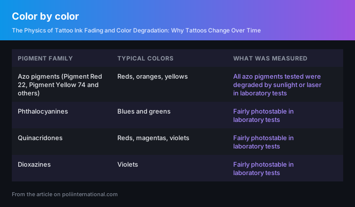
Color by color

Pigment familyTypical colorsWhat was measured
Azo pigments (Pigment Red 22, Pigment Yellow 74 and others)Reds, oranges, yellowsAll azo pigments tested were degraded by sunlight or laser in laboratory tests
PhthalocyaninesBlues and greensFairly photostable in laboratory tests
QuinacridonesReds, magentas, violetsFairly photostable in laboratory tests
DioxazinesVioletsFairly photostable in laboratory tests
Color by color
Infographic: Color by color

The table follows the review by Hauri and Hohl of the State Laboratory of Basel-City (2015). It explains the pattern tattooists see at the chair: reds and yellows from azo pigments lose strength, while phthalocyanine blues and greens hold.

Black is missing from the table because none of the studies here measured it. Black inks are usually based on carbon, not on an organic dye molecule, so the mechanism above does not apply to them in the same way.

What a tattooed person can do

Light is one of the two causes of fading, and it is the only one a person controls. Clothing over the tattoo stops the light. Sunscreen reduces the ultraviolet that reaches the skin. Neither has been tested in a study on tattoo fading, so there is no figure for how much they help; the advice follows from the mechanism, not from a trial.

The second cause, pigment leaving the skin through the body, is not something aftercare changes.

What is not known

- No study gives a fading rate per year for a tattoo on a person.
- The skin measurements cover red azo pigments. Other colors have laboratory data only.
- Nobody has measured how much sunscreen slows fading.

The breakdown products matter for another reason too. Several of the molecules that azo pigments split into are toxic or carcinogenic, which is why the same researchers raise pigment photostability as a safety question and not only a cosmetic one.

---

FAQ

Q: Why do some tattoo colors fade faster than others?
A: Because the pigments are different chemicals. In laboratory tests every azo pigment exposed to sunlight or laser broke down, and azo pigments are the usual source of red and yellow. Phthalocyanines (blue and green), quinacridones and dioxazines were fairly photostable in the same tests (Hauri and Hohl, 2015).

Q: Does sun exposure make a tattoo fade?
A: Yes, for the pigments that have been measured. Natural sunlight cleaved the red azo pigment Pigment Red 22 in solution, and in tattooed mice 32 days under a solar simulator reduced the pigment in the skin by about 60% (Engel et al., 2007 and 2010). No comparable measurement exists for every pigment, and none of these studies tested sunscreen.

Q: Where does the pigment go?
A: Part of it is broken apart by light. Part of it leaves the skin: in mice, about 32% of a red pigment was gone 42 days after tattooing without any light exposure. In samples of red tattooed human skin taken years later, researchers estimated that 87 to 99% of the original pigment was no longer there (Lehner et al., 2011).

---

Key Fact: In tattooed mice, 32 days under a solar simulator reduced the red azo pigment Pigment Red 22 in the skin by about 60% (Engel et al., Exp Dermatol 2010; PMID 19703227).

Mind map · The Physics of Tattoo Ink Fading and Color Degradation: Why Tattoos Change Over Time

  • Physics of Tattoo Ink Fading
    • Light-Induced Molecular Photodegradation

      Ultraviolet photons break chemical bonds holding organic tattoo pigment molecules together [The Physics of Tattoo Ink Fading and Color Degradation: Why Tattoos Change Over Time].

      • UV Photon Energy
      • Organic Azo Decomposition
        • Toxic Breakdown Products
    • Biological Pigment Loss

      Tattooed mice lost 32 percent of skin pigment after 42 days without light exposure [The Physics of Tattoo Ink Fading and Color Degradation: Why Tattoos Change Over Time].

      • Mouse Skin Solar Reduction
      • Human Long-Term Retention
    • Macrophage Recapture Cycle

      Dermal macrophages capture, release, and recapture tattoo pigment upon cell death in continuous cycles [The Physics of Tattoo Ink Fading and Color Degradation: Why Tattoos Change Over Time].

      • Cellular Ink Capture
      • Release Upon Cell Death
      • Recapture By New Macrophages
    • Color Photostability Variation

      Azo pigments degrade under light, whereas phthalocyanines, quinacridones, and dioxazines exhibit high photostability [The Physics of Tattoo Ink Fading and Color Degradation: Why Tattoos Change Over Time].

      • Vulnerable Red Yellow Azo
      • Resilient Blue Green Phthalocyanines
      • Stable Quinacridones and Dioxazines
    • Protective Measures and Limits

      Sunscreen and clothing mitigate light-induced fading, but aftercare cannot stop internal biological pigment clearance [The Physics of Tattoo Ink Fading and Color Degradation: Why Tattoos Change Over Time].

      • Sunscreen and Clothing Barrier
      • Uncontrollable Biological Removal
StructureProcessMaterialHazard

5 branches, 0 sources. Each branch cites the article it came from; the labels beneath it are names, not claims.

Tattoo Fading Quiz

  1. 1. What are the two primary mechanisms identified in the source material that cause tattoo pigment to diminish over time?

    • ✓ Cleavage of dye molecules by light photons and physical removal of pigment by the body — Light carries sufficient photon energy to break organic pigment chemical bonds, while cellular transport mechanisms concurrently carry pigment particles away from dermal tissue.
    • • Shedding of the epidermis through surface exfoliation and chemical breakdown by skin acidity — Tattoo pigments reside deeper within the dermal layer rather than the shedding epidermal layer, and dermal pH changes are not cited as a degradation mechanism.
    • • Thermal dissipation from dermal heat and enzymatic digestion within dermal macrophages — While macrophages engulf pigment, the body removes pigment intact rather than digesting it enzymatically, and internal skin temperature does not break pigment chemical bonds.
    • • Oxidation from ambient air exposure and mechanical displacement from daily muscular movement — Ambient oxygen does not penetrate the dermal layer to cause primary fading, nor does structural muscular action account for pigment loss.
  2. 2. Why are UVB photons carrying energy between 3.94 eV and 4.43 eV specifically capable of breaking down organic tattoo pigments?

    • ✓ This photon energy matches the energy range required to break chemical bonds holding organic pigments together — Photons within this ultraviolet energy range carry sufficient quantum energy to disrupt specific covalent bonds that give organic pigment molecules their color.
    • • This wavelength range generates localized heat that melts organic dye microcrystals in the skin — The mechanism of ultraviolet fading relies on direct photon cleavage of chemical bonds rather than photothermal melting of pigment structures.
    • • This photon energy neutralizes dermal macrophage activity, forcing cells to eject pigment particles — UV radiation directly breaks pigment molecules apart chemically rather than deactivating macrophage cellular functions.
    • • This energy level selectively dissolves heavy metal ions contained within synthetic dye formulations — The energy match described applies specifically to organic covalent chemical bonds rather than metallic ion dissolution.
  3. 3. In the study on hairless mice tattooed with Pigment Red 22 (Engel et al., 2010), how much pigment was lost after 42 days without any light exposure?

    • ✓ Approximately 32% — Quantitative measurements established that roughly one-third of the initial pigment quantity left the living skin within six weeks through body transport alone.
    • • Approximately 51% — This value represents the pigment reduction observed in the study when tattooed mouse skin was subjected to laser treatment.
    • • Approximately 60% — This percentage reflects the total pigment loss observed when tattooed mouse skin was exposed to 32 days of simulated solar light.
    • • Approximately 87% — This figure is the lower bound of estimated long-term pigment loss measured in human skin samples taken years after tattooing.
  4. 4. Based on measurements from human skin samples taken years after tattooing (Lehner et al., 2011), what estimated proportion of red azo pigment had left or decomposed in the skin?

    • ✓ 87% to 99% — Comparing years-old human tattooed skin samples (mean 0.077 mg/cm²) against fresh tattoo concentrations (0.60 to 9.42 mg/cm²) revealed that the vast majority of original pigment had vanished.
    • • 32% to 60% — This range reflects the 42-day baseline and solar simulator loss figures recorded in short-term mouse studies rather than long-term human data.
    • • 50% to 70% — This range corresponds to laser light reduction figures in short-term animal trials rather than multi-year human sample estimates.
    • • 10% to 25% — This significantly underestimates the degree of long-term pigment loss demonstrated by human skin extract measurements.
  5. 5. According to research on living skin (Baranska et al., 2018), why does a tattoo remain visibly distinct even after most of its pigment has left or decomposed?

    • ✓ Dying dermal macrophages release pigment, which is recaptured on-site by newly arriving macrophages — A continuous cycle of capture, release, and recapture by dermal macrophages holds the remaining pigment particles precisely where the needle placed them.
    • • Pigment particles bond permanently to static dermal collagen fibers that never undergo remodeling — The source material demonstrates that pigment is dynamically held within living immune cells rather than bound permanently to extracellular matrix fibers.
    • • Epidermal stem cells continually duplicate original pigment molecules during regular cellular division — Epidermal cells do not synthesize or replicate exogenous ink pigment particles.
    • • Residual pigment migrates into deep muscle tissue where light cannot penetrate or break it down — The remaining tattoo pigment stays localized within dermal macrophages rather than sinking into sub-dermal muscular layers.
  6. 6. Which family of pigments is commonly used for blue and green tattoo inks and demonstrates relative photostability in laboratory tests?

    • ✓ Phthalocyanines — Laboratory evaluations of chemical photostability confirmed that phthalocyanine structures resist breakdown under light exposure far better than azo pigments.
    • • Azo pigments — Azo pigments produce red, yellow, and orange shades and break down readily when exposed to solar or laser light.
    • • Quinacridones — While photostable, quinacridones are utilized for red, magenta, and violet hues rather than blues and greens.
    • • Dioxazines — Dioxazines are photostable compounds used primarily to produce violet shades rather than blue or green tones.
  7. 7. How do quinacridone pigments differ from azo pigments when used in red, magenta, or violet tattoo inks?

    • ✓ Quinacridones exhibit high photostability in lab tests, whereas tested azo pigments readily decompose under light — Despite producing overlapping color spectra like red, quinacridone dye molecules resist light cleavage while azo molecular bonds split under solar radiation.
    • • Quinacridones are inorganic mineral salts, whereas azo pigments consist purely of carbon black particles — Both families are organic dye classes, whereas carbon black represents a separate elemental ink category.
    • • Quinacridones are rapidly removed by body transport within weeks, whereas azo pigments remain trapped forever — Azo pigments are heavily cleared and degraded over time, and quinacridones do not exhibit accelerated bodily elimination.
    • • Quinacridones dissolve completely in human dermal fluid, whereas azo pigments remain fully insoluble — Solubility in skin fluids is not the defining distinction drawn between these two pigment families.
  8. 8. Why are black tattoo inks excluded from the comparative laboratory breakdown tables for organic dyes?

    • ✓ Black inks are typically carbon-based rather than organic dye molecules, so photodecomposition mechanisms differ — Because carbon black consists of elemental carbon particles rather than complex organic dye molecules with light-sensitive chemical bonds, the same molecular cleavage rules do not apply.
    • • Black ink particles are completely immune to bodily transport and macrophage ingestion — Macrophages handle black carbon particles in skin similarly to other inks; it is the light-driven molecular cleavage mechanism that differs.
    • • No laboratory instruments exist that can accurately quantify black pigment in biological tissues — Extraction and measurement techniques exist, but black inks were omitted from the reviewed studies because they lack organic dye structures.
    • • Black inks absorb 100% of UV radiation without undergoing any physical or structural alterations — The text notes that black ink was simply unmeasured in the cited studies due to its fundamentally non-organic chemical nature.
  9. 9. What key limitation does the source text highlight regarding recommendations to use sunscreen or protective clothing for tattoo preservation?

    • ✓ The advice follows logical optical mechanisms, but neither method has been tested in a clinical tattoo fading trial — Although light blocking prevents photon-driven chemical cleavage in theory, no empirical study has quantified the exact percentage by which sunscreen slows fading in living skin.
    • • Sunscreen chemicals react with dermal macrophages, accelerating the systemic removal of ink particles — Sunscreen acts externally on top of the skin and has not been shown to increase internal cellular transport of pigment.
    • • Protective clothing traps body heat, increasing the photothermal breakdown rate of organic pigments — Clothing effectively blocks sunlight photons without inducing thermal degradation of tattoo pigments.
    • • Sunscreen only protects against UVA radiation, leaving pigment vulnerable to high-energy UVB photons — The limitation emphasized is the complete lack of empirical testing data rather than a specific spectral failure of sunscreen.
  10. 10. Besides cosmetic fading, why is the light-induced breakdown of azo pigments considered an important safety issue?

    • ✓ Several breakdown products resulting from azo pigment cleavage are toxic or carcinogenic — Splitting azo chemical bonds creates smaller cleavage molecules, many of which pose systemic toxicity or cancer risks to the host body.
    • • Cleaved azo fragments aggregate into crystalline deposits that permanently block lymphatic drainage — The primary safety concern highlighted is the toxicological and carcinogenic nature of molecular breakdown products, not physical lymphatic blockade.
    • • Azo photodecomposition triggers immediate systemic autoimmune destruction of healthy skin tissues — The cited health risk relates to chemical toxicity and carcinogenicity of split products rather than an autoimmune disease state.
    • • Light exposure turns azo pigments into radioactive isotopes within the dermal tissue layer — Light cleavage involves breaking molecular bonds between atoms, not nuclear alterations that generate radiation.

Extended Reference Table

8 of 8 rows

Extended reference table generated from the article and its verified research sources. Bracketed numbers jump to the matching entry in the Technical References below.

SafetyItem / Practice / Material Category Applicable Standards or Guidelines Biological or Technical Mechanism
✅Pigment Blue 15 (C.I. 74160)Organic Colour PigmentResolution ResAP(2008)1 / Swiss Ordinance 817.023.41Acts as a copper phthalocyanine blue colourant; suspended as insoluble particles in ink matrix, detectable by characteristic ion cluster [M+H]+ at m/z 575.
✅Pigment Green 7 (C.I. 74260)Organic Colour Pigment (Prohibited)Resolution ResAP(2008)1 / Swiss Ordinance 817.023.41Chlorinated copper phthalocyanine green pigment; banned under regulations due to safety/toxicological concerns or missing safety data, absorbed as insoluble suspended particles in skin dermis.
✅Pigment Red 170 (C.I. 12475)Organic Colour PigmentResolution ResAP(2008)1 / Swiss Ordinance 817.023.41Naphthol AS red pigment; yields characteristic ions [M+H]+ (m/z 455), [M+Na]+ (m/z 477), and fragment ion m/z 318 upon laser desorption ionisation.
✅Pigment Red 254 (C.I. 56110)Organic Colour PigmentResolution ResAP(2008)1 / Swiss Ordinance 817.023.41Diketopyrrolopyrrole red pigment; demonstrates strong ionisation potential allowing detection at low concentrations (LOD ~1% w/w).
✅Pigment Red 122 (C.I. 73915)Organic Colour Pigment (Prohibited)Resolution ResAP(2008)1 / Swiss Ordinance 817.023.41Quinacridone magenta pigment; banned organic colourant forming characteristic ions [M+H]+ (m/z 341) and [M+Na]+ (m/z 363).
✅Pigment Violet 23 (C.I. 51319)Organic Colour Pigment (Prohibited)Resolution ResAP(2008)1 / Swiss Ordinance 817.023.41Dioxazine violet pigment; banned under European regulations, produces molecular ion cluster [M]+ at m/z 588 and fragment ion clusters at m/z 554 and m/z 520.
✅Pigment Yellow 12 (C.I. 21090)Organic Colour PigmentResolution ResAP(2008)1 / Swiss Ordinance 817.023.413,3'-Dichlorobenzidine-based diarylide yellow pigment; exhibits weak molecular ion signals under LDI but enhanced molecular ion intensity ([M]+ at m/z 628) when combined with 2,5-dihydroxybenzoic acid (DHB) matrix.
✅Pigment Green 36 (C.I. 74265)Organic Colour PigmentResolution ResAP(2008)1 / Swiss Ordinance 817.023.41Brominated copper phthalocyanine green pigment; susceptible to mass signal suppression by non-ionic surfactants (e.g., Triton X-100), requiring ethanolic sample clean-up for accurate detection.

Free Body Art Wall Posters

Print-ready reference posters for your studio or your own wall. Free to download and display, embed on your own site with attribution. Click any poster to preview it full size before downloading.

Choose and download all 40 posters

Technical_References_Archive

  • [1]Engel E, Vasold R, Santarelli F, et al. Tattooing of skin results in transportation and light-induced decomposition of tattoo pigments: a first quantification in vivo using a mouse model. Exp Dermatol. 2010;19(1):54-60. PMID 19703227.
  • [2]Lehner K, Santarelli F, Penning R, et al. The decrease of pigment concentration in red tattooed skin years after tattooing. J Eur Acad Dermatol Venereol. 2011;25(11):1340-1345. PMID 21349116.
  • [3]Engel E, Spannberger A, Vasold R, et al. Photochemical cleavage of a tattoo pigment by UVB radiation or natural sunlight. J Dtsch Dermatol Ges. 2007;5(7):583-589. PMID 17610608.
  • [4]Cui Y, Spann AP, Couch LH, et al. Photodecomposition of Pigment Yellow 74, a pigment used in tattoo inks. Photochem Photobiol. 2004;80(2):175-184. PMID 15362942.
  • [5]Hauri U, Hohl C. Photostability and breakdown products of pigments currently used in tattoo inks. Curr Probl Dermatol. 2015;48:164-169. PMID 25833639.
  • [6]Baranska A, Shawket A, Jouve M, et al. Unveiling skin macrophage dynamics explains both tattoo persistence and strenuous removal. J Exp Med. 2018;215(4):1115-1133. PMID 29511065.

About the Author

PP

Patrick Poli

Chief Engineer, Poli International · Creator of BioFlex® body jewelry

Patrick has spent 25+ years manufacturing body jewellery and studio equipment, and is the creator of BioFlex® body jewelry. He created the push-fit system so jewellery separates on impact instead of tearing the piercing. He also reworked the push-fit discs in medical-grade soft polymer, EO-sterilised, for people fighting bumps and keloids.

More about Patrick →

Last reviewed: 2026-10-12 · Published: 2026-10-11

Related Tools & Reading

Continue Reading