Shop
Clinical Physics

Red Light Therapy on Tattoos and Piercings: What Photobiomodulation Really Delivers

PP

Chief Engineer

Patrick Poli

Journal Date

2026-09-29

Last Reviewed

2026-10-06

Executive Summary. Red light therapy on a new tattoo or piercing is photobiomodulation: photons absorbed by mitochondrial chromophores at doses far too low to heat tissue. On intact skin, 600 to 900 nm light reaches roughly one to six millimetres depending on wavelength, beam width and melanin, which puts a piercing fistula inside the therapeutic window and tattoo pigment at its edge. The physics is defensible; the tattoo-specific human evidence is not.

Photobiomodulation Is Absorption, Not Heat

Photobiomodulation (PBM), the term that replaced low-level laser therapy, is a non-thermal interaction. Photons are absorbed by chromophores in the mitochondrial respiratory chain, principally cytochrome c oxidase, and the absorbed energy shifts signalling rather than temperature: nitric oxide release, a transient rise in reactive oxygen species, and downstream transcription of genes involved in proliferation and migration. de Freitas and Hamblin set out the mechanism in 2016, and the 2024 American Academy of Dermatology review by Maghfour and colleagues restates it with one constraint attached: the effect is dose-dependent and disappears outside a narrow band.

This is not the regime of laser tattoo removal. Both use red and near-infrared light, and the wavelength tables look similar, but photothermolysis relies on preferential absorption by pigment followed by heat diffusion that denatures the target, while PBM relies on sub-thermal absorption that leaves tissue intact. A device that clears ink will cook a healing fistula. A device that is safe on a healing fistula will never touch ink.

How Deep the Light Actually Gets

Skin scatters. Collagen bundles and cell membranes deflect photons repeatedly, so a beam fans out within the first millimetre and loses intensity with depth. Absorption on top of that scattering is dominated by haemoglobin below about 600 nm, water above roughly 1100 nm, and melanin throughout, which is why the therapeutic window sits between 600 and 900 nm. Tattoo pigment sits at 1 to 2 mm, in the papillary and upper reticular dermis, as covered in dermal depth and skin histology.

Ash and colleagues modelled light-tissue interaction in 2017 and reported two clinically useful findings. Penetration increases with wavelength across the window, so 810 nm reaches deeper than 660 nm, and it increases with beam width, because a wider beam loses proportionally less to lateral divergence. Their model puts therapeutic devices at a few millimetres of meaningful dose, not centimetres.

Melanin removes dose before it arrives. In deeper skin tones more photons are absorbed in the epidermis, so the same setting, distance and timer deliver less energy to the dermis than they do in lighter skin. The optical behaviour behind that is covered in skin optics and pigment appearance.

Dose Is Not Brightness

Fluence equals irradiance multiplied by time. A device delivering 100 mW/cm2 for five minutes doses 30 J/cm2. Double the time and you double the fluence. Double the distance and you may quarter it. Panels sold for home use are usually specified in lux or electrical wattage, and neither translates into irradiance at the skin: lux is weighted for human photopic vision and says little about 660 nm photon flux. The honest specification is milliwatts per square centimetre at the surface, measured with a power meter, and the honest treatment record is joules per square centimetre delivered.

Dose does not scale linearly with benefit either. Huang and colleagues described a biphasic dose response in 2011: too little light produces no measurable effect, an optimum range produces the response, and doses above it suppress the response. Where a home panel appears to do nothing, the usual cause is underdosing at the surface. Where it irritates, the usual cause is a session long enough to add a thermal component.

Parameter reporting compounds the problem. Cronshaw and colleagues reviewed PBM dose parameters in 2020 and found wavelength, irradiance, fluence, pulse structure and treatment interval reported inconsistently across studies, which is a large part of why meta-analyses disagree. Mosca and colleagues made the same point for wound care in 2019 and proposed a reporting framework so the field could compare itself.

What the Evidence Shows for Tattoos and Piercings

Direct tattoo evidence is close to absent. A PubMed search for photobiomodulation in tattoo treatment returns one relevant study: Imrigha and colleagues treated wounds in a rat model after Q-switched Nd:YAG tattoo removal and reported improved wound outcomes. That is one preclinical study in animals, with no human replication. Nothing indexed in PubMed tests PBM on human tattoo healing.

Evidence one step out is better but still not tattoo-specific. Deana and colleagues meta-analysed PBM in burn wound healing in 2021 and found the pooled evidence came entirely from preclinical studies. Taha and colleagues meta-analysed low-level laser therapy for healing and pain in skin wounds in 2024 and reported probable benefit with substantial heterogeneity in dose and device. The strongest human evidence for PBM sits in oral mucositis, diabetic foot ulcers and post-surgical wounds. None of those is a puncture wound through a cartilage plate with a foreign body seated in it.

ClaimPhysical plausibilityHuman evidence
Faster healing of a fresh tattooWeak: dermal pigment sits at 1 to 2 mm and the epidermis absorbs dose on the way inNone indexed. One rat study after laser removal
Less pain at a healing piercingPlausible: the fistula wall is within the millimetres that receive doseNone in piercings. Indirect evidence from skin wounds
Less redness and swelling after a sessionPlausible through transient vasoactive effectsIndirect, from burn and post-surgical wound studies
Faster ink clearance after laser sessionsWeak: the wavelength and fluence windows do not overlap with photothermolysisPreclinical only, wound outcomes rather than ink outcomes
Safer healing in darker skin tonesWeak: epidermal melanin absorbs more of the incident doseNot studied. PBM dosing is rarely reported by skin tone

What to Do With That

1. Ask for irradiance in milliwatts per square centimetre and calculate fluence yourself. Ten minutes on a bright panel is not a dose.
2. Do not treat an infected or open wound with light instead of assessment. PBM is not antimicrobial, and infection after a tattoo or piercing needs clinical review.
3. On a fresh tattoo, wait until the epidermis has closed, roughly two weeks, then treat intact skin only. In the first fortnight the wound environment matters more, as covered in what belongs on a new tattoo and the aftercare science overview.
4. On piercings the physics is more favourable, because the fistula is shallow. Remove or displace metal jewelry where you can, since polished metal reflects and the shadow it casts is real.
5. Track the wound, not the session. Photographs at a fixed distance under the same light beat a diary of minutes, and the healing tracker and aftercare schedule generator record it properly.
6. Keep client expectations honest. If a healing tattoo is not settling, the useful question is what is mechanically or microbiologically wrong with it, not how many more joules of red light it can absorb. The phases it has to pass through are set out in wound healing biology, and the adjacent non-thermal option is covered in cold atmospheric plasma.
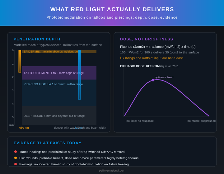
Red light therapy on tattoos and piercings: penetration depth, fluence and evidence quality
Red light therapy on tattoos and piercings: what reaches the target, at what dose, and with what evidence.


FAQ


Can red light therapy help heal a new tattoo?


There is a plausible mechanism, but the tattoo-specific human evidence does not yet support a healing claim. Photobiomodulation at 600 to 900 nm reaches the papillary and upper reticular dermis where tattoo pigment sits, and cytochrome c oxidase absorption can shift signalling toward proliferation and migration, but Maghfour and colleagues' 2024 American Academy of Dermatology review stresses the effect is dose-dependent and vanishes outside a narrow band. No robust controlled trial has shown faster or cleaner tattoo healing from PBM, so treat any benefit as theoretical until better data arrive.


Is red light therapy safe on a healing piercing?


It is likely the safer of the two applications because a piercing fistula sits within the 1 to 6 mm penetration depth that 600 to 900 nm light achieves on intact skin. The risk is not the wavelength but the dose: a session long enough to add a thermal component can irritate a fresh fistula, and Huang and colleagues described a biphasic dose response in 2011 where too little does nothing and too much suppresses the response. Keep sessions sub-thermal and verify irradiance at the surface rather than trusting lux or wattage ratings.


Does red light therapy remove or fade tattoo ink?


No, and confusing the two modalities is dangerous. Laser tattoo removal works by photothermolysis, where pigment absorbs light preferentially and heat diffusion denatures the target, while PBM relies on sub-thermal absorption that leaves tissue intact. As the article notes, a device that clears ink will cook a healing fistula, and a device that is safe on a healing fistula will never touch ink.


How deep does red light actually penetrate into skin?


Ash and colleagues modelled light-tissue interaction in 2017 and put therapeutic devices at a few millimetres of meaningful dose, not centimetres. Penetration increases with wavelength across the window, so 810 nm reaches deeper than 660 nm, and it also increases with beam width because a wider beam loses proportionally less to lateral divergence. Melanin removes dose before it arrives, so the same setting, distance and timer deliver less energy to the dermis in deeper skin tones.


How do I know if my red light device is delivering enough dose?

Fluence equals irradiance multiplied by time, so a device delivering 100 mW/cm² for five minutes doses 30 J/cm². Panels sold for home use are usually specified in lux or electrical wattage, and neither translates into irradiance at the skin because lux is weighted for human photopic vision and says little about 660 nm photon flux. The honest specification is milliwatts per square centimetre at the surface measured with a power meter, and the honest treatment record is joules per square centimetre delivered.


Mind map · Red Light Therapy on Tattoos and Piercings: What Photobiomodulation Really Delivers

  • Photobiomodulation for Body Art
    • Non-Thermal Cellular Mechanism

      Cytochrome c oxidase absorbs photons to shift signaling without heating tissue [Red Light Therapy on Tattoos and Piercings: What Photobiomodulation Really Delivers].

      • Cytochrome C Oxidase
        • Nitric Oxide Release
      • Sub-Thermal Light Absorption
        • Preserves Intact Tissue
    • Optical Window Penetration

      Red and near-infrared light reaches one to six millimetres inside skin [Red Light Therapy on Tattoos and Piercings: What Photobiomodulation Really Delivers].

      • Dermal Penetration Limits
        • 810 nm Reaches Deeper
      • Epidermal Melanin Absorption
        • Melanin Reduces Dermal Energy
    • Biphasic Dose Response

      Photobiomodulation exhibits a biphasic response where excessive dose suppresses biological activity [Red Light Therapy on Tattoos and Piercings: What Photobiomodulation Really Delivers].

      • Fluence Calculation Method
        • Irradiance Multiplied By Time
      • Overdosing Risk
    • Human Evidence Gap

      Zero PubMed indexed clinical trials test photobiomodulation on fresh human tattoos [Red Light Therapy on Tattoos and Piercings: What Photobiomodulation Really Delivers].

      • Preclinical Animal Study
        • Laser Tattoo Removal Model
      • Indirect Wound Evidence
    • Practical Application Rules

      Fresh tattoos require waiting two weeks for epidermal closure before light therapy [Red Light Therapy on Tattoos and Piercings: What Photobiomodulation Really Delivers].

      • Epidermal Closure Wait
      • Infection Clinical Assessment
StructureProcessMaterialHazard

5 branches, 0 sources. Each branch cites the article it came from; the labels beneath it are names, not claims.

Photobiomodulation Quiz

  1. 1. What is the primary cellular mechanism behind photobiomodulation (PBM)?

    • • Selective photothermolysis that thermally denatures target tissue pigment. — This describes the mechanism of laser tattoo removal, which relies on heat energy to denature target structures rather than PBM.
    • ✓ Sub-thermal photon absorption by mitochondrial chromophores like cytochrome c oxidase. — PBM relies on photons being absorbed by mitochondrial enzymes to shift cell signaling without increasing tissue temperature.
    • • Thermal disruption of cell membranes via high-intensity light absorption. — PBM is a non-thermal process operating at low energy levels that leave cell membranes and tissue structures intact.
    • • Direct photo-oxidation of dermal extracellular matrix collagen fibers. — PBM specifically targets respiratory enzymes within mitochondria rather than directly breaking down extracellular collagen.
  2. 2. How does photobiomodulation (PBM) differ physically from laser tattoo removal?

    • • PBM uses ultra-short pulses to shatter pigment, whereas laser removal uses continuous heat. — Laser removal shatters pigment through thermal mechanism, whereas PBM does not shatter pigment at all.
    • • PBM operates in the ultraviolet spectrum, whereas laser removal operates in the near-infrared spectrum. — Both PBM and laser tattoo removal utilize red and near-infrared wavelengths.
    • ✓ PBM relies on sub-thermal absorption to keep tissue intact, whereas laser removal uses heat diffusion to denature targets. — PBM uses light doses far too low to heat tissue, whereas laser removal relies on thermal absorption to break down pigment.
    • • PBM delivers significantly higher fluence levels than laser removal to reach deeper dermal layers. — Laser removal utilizes higher energy density to shatter ink, whereas PBM requires low fluence within a narrow non-thermal band.
  3. 3. According to optical modeling in skin tissue, which parameters increase the penetration depth of light within the therapeutic window (600 to 900 nm)?

    • • Lowering the wavelength and narrowing the light beam width. — Shorter wavelengths undergo higher attenuation, and narrower beams suffer higher photon loss from lateral divergence.
    • ✓ Increasing the wavelength and widening the light beam width. — Longer wavelengths within the window scatter less in tissue, and wider beams lose proportionally less energy to lateral divergence.
    • • Increasing light irradiance while narrowing the light beam width. — While irradiance increases surface power, beam width is what dictates the rate of lateral photon divergence loss.
    • • Shifting the wavelength to the ultraviolet range while increasing total session fluence. — Ultraviolet light is rapidly absorbed in upper epidermal layers and cannot penetrate into the deep dermis.
  4. 4. What impact does a higher concentration of epidermal melanin have on photobiomodulation dosing?

    • • It acts as an optical reflector, increasing the effective light dose delivered to the dermis. — Melanin absorbs incoming photons in the outer skin layer rather than reflecting them into deeper tissues.
    • ✓ It absorbs photons in the epidermis, reducing the light energy delivered to the underlying dermis. — Higher epidermal melanin absorbs a greater portion of incident light, leaving fewer photons to reach target chromophores in the dermis.
    • • It converts red light into therapeutic heat, accelerating cellular migration in dermal layers. — PBM mechanisms are sub-thermal, and photon absorption by epidermal melanin diminishes the desired dermal dose.
    • • It expands the therapeutic window, allowing shorter wavelengths to reach deeper skin structures. — Melanin absorbs light across the spectrum and reduces depth penetration regardless of the wavelength used.
  5. 5. What defines the biphasic dose response associated with photobiomodulation?

    • • Biological benefits scale infinitely as treatment time and panel brightness increase. — PBM does not scale linearly, and exceeding optimal parameters leads to a suppression of cellular response.
    • ✓ Doses below a threshold produce no effect, an optimal range gives benefit, and higher doses suppress the response. — The biphasic dose response curve demonstrates that both underdosing and overdosing fail to achieve therapeutic stimulation.
    • • Low irradiance causes thermal tissue damage, whereas high irradiance produces purely photochemical signaling. — Thermal effects and irritation occur at excessively high doses or long sessions, not at low irradiance.
    • • Pulsed light inhibits mitochondrial activity, whereas continuous light output reliably stimulates it. — The biphasic response relates to the total energy density delivered rather than strictly the pulse structure.
  6. 6. Why are commercial light panels rated in lux insufficient for calculating a clinical photobiomodulation dose?

    • • Lux measures thermal heat output rather than electrical energy consumed by light panels. — Lux measures visible light brightness relative to human eye perception, not thermal energy.
    • ✓ Lux is weighted for human visual perception and does not accurately report red photon flux or irradiance. — Lux measures photopic human vision sensitivity rather than the milliwatts per square centimeter required to measure therapeutic light dose.
    • • Lux overestimates near-infrared light energy while completely ignoring visible spectrum light. — Lux is heavily weighted toward green visible light and does not measure near-infrared wavelengths.
    • • Lux is a measurement unit that applies exclusively to coherent laser beams rather than LED light panels. — Lux can evaluate any broad visual light source, but it lacks physical relevance for therapeutic photon flux.
  7. 7. What does the currently indexed scientific literature demonstrate regarding photobiomodulation on human tattoo healing?

    • • Multiple clinical trials confirm significantly accelerated wound recovery for new human tattoos. — There are zero indexed human clinical trials evaluating PBM on fresh tattoo healing.
    • ✓ A single preclinical rat study evaluated wound outcomes after laser removal, with no human replication. — The direct evidence is limited to one animal study in rats following Nd:YAG laser treatment, without human clinical studies.
    • • Human studies show reliable accelerated ink clearance when PBM is paired with Q-switched laser sessions. — The single existing animal study evaluated wound outcomes post-laser removal, not ink clearance performance in humans.
    • • Observational studies demonstrate that PBM prevents hyperpigmentation and keloiding in fresh tattoos. — PBM clinical outcomes on tattoo scarring and hyperpigmentation have not been tested in human trials.
  8. 8. Why is a healing piercing fistula a more physically plausible target for photobiomodulation than tattoo pigment?

    • • Piercing fistulas lack blood vessels, completely preventing absorption by haemoglobin. — Fistula walls are vascular tissue where haemoglobin absorption still takes place.
    • ✓ The piercing fistula sits shallowly within the millimetres that receive a therapeutic light dose. — Because light penetrates a few millimetres into tissue, a shallow piercing fistula sits cleanly inside the therapeutic depth window.
    • • Metal jewelry acts as a conductor that absorbs red photons and re-radiates them into surrounding cartilage. — Metal jewelry reflects light photons away and creates a shadow that obstructs light delivery.
    • • Cartilage tissue converts near-infrared energy into a mild heat shock response that speeds up collagen synthesis. — PBM acts through non-thermal mitochondrial pathways rather than thermal stimulation of cartilage.
  9. 9. What protocol is recommended regarding photobiomodulation timing on a freshly applied tattoo?

    • • Apply high-irradiance light immediately following the tattoo session to seal broken dermal blood vessels. — Open tattoo wounds require proper surface wound care rather than immediate light panel exposure.
    • ✓ Wait roughly two weeks until the epidermis has fully closed before treating intact skin. — During the first fortnight, maintaining a healthy wound environment is paramount; light therapy should wait until skin is intact.
    • • Use red light panels daily during the first 48 hours to sanitize open puncture wounds. — PBM is a non-thermal signaling process and has no direct antimicrobial action against wound infections.
    • • Double the calculated fluence during the initial week to overcome open epidermal scattering. — Increasing fluence on open wound tissue risks thermal irritation and exceeds optimal therapeutic dosing windows.
  10. 10. What physical preparation is advised when delivering photobiomodulation to a healing body piercing?

    • • Keep polished metal jewelry in place so it reflects light back into the deeper fistula walls. — Polished metal reflects incident light away from the tissue and casts an unexposed shadow beneath it.
    • ✓ Remove or displace metal jewelry whenever possible to avoid reflection and shadowing over target tissue. — Metal jewelry reflects photons and casts shadows, preventing the shallow target tissue behind it from receiving the intended dose.
    • • Preheat the jewelry using thermal light to create localized thermal photothermolysis inside the fistula. — Thermal heating causes tissue denaturation, directly contradicting the non-thermal principle of PBM.
    • • Apply a thick coat of petroleum jelly around the jewelry to refract light deeper into cartilage. — Topical barriers and metal shadowing obstruct light delivery and alter unpredictable dose distribution.

Extended Reference Table

8 of 8 rows

Extended reference table generated from the article and its verified research sources. Bracketed numbers jump to the matching entry in the Technical References below.

SafetyItem or Practice Category Wavelength or Spectrum Range Biological Mechanism Target Tissue Depth Relevant Standard or Regulator
✅Red Light PhotobiomodulationPhotobiomodulation / Low-Level Light Therapy630–660 nmStimulates mitochondrial ATP production, supports collagen synthesis, reduces inflammation, and improves circulationSkin tissue / Cellular levelFDA
✅Near-Infrared PhotobiomodulationPhotobiomodulation / Low-Level Light Therapy810–850 nmStimulates cellular function and ATP production without targeting or fragmenting pigmentDeep tissue / Underlying muscleFDA
✅Arndt-Schulz Biphasic Dose Response ProtocolClinical Photobiomodulation Protocol / Dosing Standard600nm to 1100nmBiphasic response: low-to-moderate doses stimulate cellular processes; excessive doses (>50-100 J/cm²) inhibit cellular function and trigger localized inflammationVariable (Dermis to Deep Joint/Brain)Arndt-Schulz Law (Scientific Dose Model)
✅TattooingBody Modification / Skin Injury-Needle punctures deposit pigment into the dermis triggering inflammation, repair, and remodelingDermisAmerican Academy of Dermatology
✅Lumaflex Red Light Therapy DevicesMedical Device / Home Healthcare DeviceRed and near-infrared lightInfluences mitochondrial signaling, cellular activity, and inflammatory pathways for tissue recovery-FDA
✅Standard Tattoo AftercareClinical Practice / Care Protocol-Protects recovering skin, prevents infection, and allows natural repair processes to occurEpidermis and DermisAmerican Academy of Dermatology
✅Laser Tattoo RemovalHigh-Intensity Laser Procedure / Photothermolysis / Dermatological procedureWavelengths tuned specifically to pigment absorption (e.g., Red and near-infrared light / Q-switched Nd:YAG)Generates high-intensity photomechanical force to shatter and fragment ink particles; preferential light absorption by ink pigment causing heat diffusion that denatures the targetDermis layer (1 to 2 mm; papillary and upper reticular dermis)-
✅Ultraviolet B (UVB) ExposureUltraviolet Light TherapyUVB spectrumProduces vitamin D3 in human skin (can damage DNA/burn skin unlike RLT)Epidermis / Skin-

Free Body Art Wall Posters

Print-ready reference posters for your studio or your own wall. Free to download and display, embed on your own site with attribution. Click any poster to preview it full size before downloading.

Choose and download all 40 posters

Technical_References_Archive

  • [1]Imrigha NAA, Bidin N, Lau PS, Musa N, Zakaria N, Krishnan G. (2017). Photobiomodulation therapy on wound treatment subsequent to Q-switched Nd: YAG laser tattoo removal in rat model. J Biophotonics, 10(10), 1287-1291. PMID 28464516. [PMID 28464516](https://pubmed.ncbi.nlm.nih.gov/28464516/)
  • [2]Maghfour J, Ozog DM, Mineroff J, Jagdeo J, Kohli I, Lim HW. (2024). Photobiomodulation CME part I: Overview and mechanism of action. J Am Acad Dermatol, 91(5), 793-802. PMID 38309304. [PMID 38309304](https://pubmed.ncbi.nlm.nih.gov/38309304/)
  • [3]de Freitas LF, Hamblin MR. (2016). Proposed Mechanisms of Photobiomodulation or Low-Level Light Therapy. IEEE J Sel Top Quantum Electron, 22(3), 7000417. PMID 28070154. [PMID 28070154](https://pubmed.ncbi.nlm.nih.gov/28070154/)
  • [4]Ash C, Dubec M, Donne K, Bashford T. (2017). Effect of wavelength and beam width on penetration in light-tissue interaction using computational methods. Lasers Med Sci, 32(8), 1909-1918. PMID 28900751. [PMID 28900751](https://pubmed.ncbi.nlm.nih.gov/28900751/)
  • [5]Huang YY, Sharma SK, Carroll J, Hamblin MR. (2011). Biphasic dose response in low level light therapy - an update. Dose Response, 9(4), 602-618. PMID 22461763. [PMID 22461763](https://pubmed.ncbi.nlm.nih.gov/22461763/)
  • [6]Mosca RC, Ong AA, Albasha O, Bass K, Arany P. (2019). Photobiomodulation Therapy for Wound Care: A Potent, Noninvasive, Photoceutical Approach. Adv Skin Wound Care, 32(4), 157-167. PMID 30889017. [PMID 30889017](https://pubmed.ncbi.nlm.nih.gov/30889017/)
  • [7]Cronshaw M, Parker S, Anagnostaki E, Mylona V, Lynch E, Grootveld M. (2020). Photobiomodulation Dose Parameters in Dentistry: A Systematic Review and Meta-Analysis. Dent J (Basel), 8(4), 114. PMID 33036145. [PMID 33036145](https://pubmed.ncbi.nlm.nih.gov/33036145/)
  • [8]Deana NF, Alves N, Zaror C, Del Sol M, Bagnato VS. (2021). Photobiomodulation Therapy in Burn Wound Healing: Systematic Review and Meta-Analysis of Preclinical Studies. Photobiomodul Photomed Laser Surg, 39(7), 439-452. PMID 34264767. [PMID 34264767](https://pubmed.ncbi.nlm.nih.gov/34264767/)
  • [9]Taha N, Daoud H, Malik T, Shettysowkoor J, Rahman S. (2024). The Effects of Low-Level Laser Therapy on Wound Healing and Pain Management in Skin Wounds: A Systematic Review and Meta-Analysis. Cureus, 16(10), e72542. PMID 39610644. [PMID 39610644](https://pubmed.ncbi.nlm.nih.gov/39610644/)

About the Author

PP

Patrick Poli

Chief Engineer, Poli International · Creator of BioFlex® body jewelry

Patrick has spent 25+ years manufacturing body jewellery and studio equipment, and is the creator of BioFlex® body jewelry. He created the push-fit system so jewellery separates on impact instead of tearing the piercing. He also reworked the push-fit discs in medical-grade soft polymer, EO-sterilised, for people fighting bumps and keloids.

More about Patrick →

Last reviewed: 2026-10-06 · Published: 2026-09-29

Related Tools & Reading

Continue Reading