
How Ink Reads Through Skin Across Fitzpatrick Types

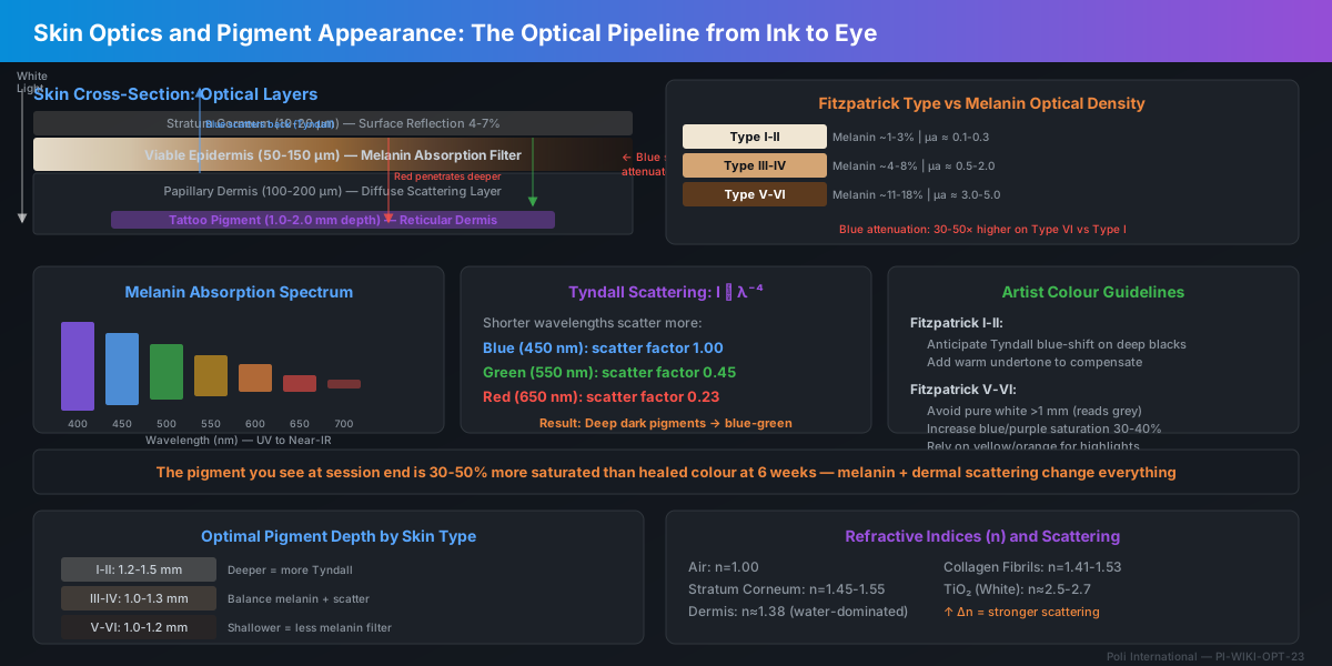
Tattoo pigment sits in the dermis beneath a living optical filter: the epidermis with its melanin content. Melanin absorbs strongly in the UV and blue regions (300-500 nm) and decreases through the visible spectrum, meaning that on darker skin, blue and violet pigments are preferentially attenuated. Dermal collagen scatters light diffusely, creating the Tyndall effect that shifts deeply-placed dark pigments toward blue-green. Understanding these optical principles allows artists to predict healed colour across Fitzpatrick types I-VI and choose inks that read true through each client's skin.
⚡ Quick Reference
Critical Numbers
- Epidermal thickness50-150 μm (palms/soles up to 600 μm); melanin concentrated in basal layer at 50-100 μm depth
- Dermal papillae depth100-200 μm, tattoo ink deposited at 1-2 mm in the reticular dermis, beneath the melanin filter
- Melanin absorption peak300-350 nm (UV); declines ~10x from 400 nm (violet) to 700 nm (red), blue end attenuated more
- Fitzpatrick I melanin content~1.3% epidermal volume; Fitzpatrick VI: ~11-18%, up to 14x difference in optical density
- Tyndall scattering wavelength dependenceI ∝ λ⁻⁴, blue light (450 nm) scatters ~5x more than red light (650 nm) in dermal collagen
- Dermal reduced scattering coefficient (μs')1-5 mm⁻¹ at 633 nm, varying with collagen density and hydration
- Epidermal melanin absorption coefficient (μa)0.1-5 mm⁻¹ across Fitzpatrick I-VI at 500 nm
- Colour shift on Fitzpatrick V-VI vs I-IIreds appear ~30% darker; yellows lose contrast; whites become grey at depth >1.5 mm
- Optimal pigment depth for colour fidelity1.0-1.5 mm, shallower risks epidermal migration; deeper adds blue-shift from Tyndall scattering
- Fresh vs healedepidermis regenerates over ink in 4-6 weeks, adding the melanin filter; healed colour is always 15-25% less saturated than fresh
Optical parameters governing how tattoo pigment is perceived through skin. These values determine colour shift, contrast, and longevity across skin types.
When a client looks at their healed tattoo, they are not looking at ink. They are looking at light that has travelled through the epidermis, scattered through the dermal collagen matrix, reflected off pigment particles, and travelled back out through the same optical path. Every step in that journey modifies the colour. The artist who understands this optical pipeline can predict the final result. The one who does not is guessing.
Skin as an Optical Medium
Skin is a turbid, multi-layered medium with wavelength-dependent absorption and scattering. The stratum corneum (10-20 μm) is a low-scattering keratin layer that contributes surface reflection (~4-7% of incident light). The viable epidermis (50-100 μm) contains melanin in keratinocytes and melanocytes, functioning as a broadband optical attenuator strongest in the UV-blue region. The dermis (1-4 mm) is dominated by collagen fibrils (0.1-10 μm diameter) that produce Mie and Rayleigh scattering. Tattoo pigment sits in fibroblasts and macrophages within the reticular dermis at 1-2 mm depth, beneath both the melanin filter and the most turbid scattering layer. The optical path to the ink and back is typically 2-4 mm of living tissue.
Melanin: The Biological Optical Filter
Melanin is a broadband absorber with an absorption spectrum that decreases quasi-exponentially from the UV through the visible to the near-infrared. At 400 nm (violet/blue), eumelanin absorption is approximately 10× higher than at 700 nm (red). This means that on Fitzpatrick V-VI skin, the epidermal melanin layer acts as a graduated blue filter: violet and blue wavelengths are strongly attenuated before they ever reach the dermal ink, while red and near-infrared pass through with relatively little loss. The practical consequence is that blue, purple, and cool-toned pigments read significantly darker and less saturated on darker skin, while warm pigments (red, orange, yellow) retain more of their chroma. Pheomelanin (red/yellow melanin found in higher proportion in Fitzpatrick I-II) absorbs more in the green-yellow region, contributing to the characteristic warm undertone of lighter skin.
Dermal Scattering and the Tyndall Effect
The dermis is a dense network of collagen fibrils with diameters of 20-100 nm (individual fibrils) to 1-10 μm (fibre bundles). These structures are comparable to or larger than visible wavelengths (400-700 nm), producing a combination of Rayleigh scattering (particles << λ, I ∝ λ⁻⁴) and Mie scattering (particles ~ λ). The λ⁻⁴ dependence means that blue light (450 nm) scatters approximately 5 times more strongly than red light (650 nm) in the dermal matrix. This is the Tyndall effect in skin: when white light enters the dermis, blue wavelengths are preferentially scattered back toward the surface before reaching deeper structures, while red wavelengths penetrate further. A dark pigment particle at 1.5-2.0 mm depth is illuminated primarily by the red-shifted light that survived the journey down, and the light scattered back is again blue-depleted. The net result: deep black ink appears blue-green through the overlying dermis. This is not a pigment chemistry problem, it is pure optics. The same black ink placed at 0.5 mm depth in a shallow area reads true black; at 2 mm it reads navy.
Patrick's Note
"Thomas Fitzpatrick's 1975 classification (Types I-VI) was originally developed to quantify UV sensitivity for phototherapy dosing. It has since become the clinical standard for skin phototype characterisation. Type I (always burns, never tans: pale white, red hair common) corresponds to minimal eumelanin, epidermal melanin volume fraction ~1-2%, optical density at 500 nm ~0.1 mm⁻¹. Type VI (never burns, deeply pigmented: dark brown) corresponds to maximal eumelanin, volume fraction ~11-18%, optical density at 500 nm ~3-5 mm⁻¹. Between Types I and VI, the epidermal optical filter increases in density by a factor of 30-50× at blue wavelengths and 10-15× at red wavelengths. The Fitzpatrick scale is not linear: the optical density jump from Type III to IV is larger than from I to II or V to VI, corresponding to the clinical observation that medium-toned skin shows the most variable healed tattoo outcomes."
Founder & Piercing Expert
Poli International
Pigment Depth and Perceived Colour
Tattoo pigment is deposited at 1-2 mm in the reticular dermis. The exact depth varies with anatomical site (thinner dermis on face/neck: 0.8-1.2 mm; thicker on back/thigh: 1.5-2.5 mm) and artist technique. Depth affects perceived colour through two mechanisms: (1) deeper pigment is behind more melanin (epidermal attenuation), and (2) deeper pigment is behind more dermal collagen (Tyndall scattering blue-shift). For Fitzpatrick I-II, the Tyndall effect dominates: deeper pigment appears cooler/bluer. For Fitzpatrick V-VI, melanin absorption dominates: deeper pigment appears darker and warmer-toned (blue end filtered out). The optimal compromise depth for colour fidelity across all skin types is 1.0-1.5 mm, balancing melanin path length against Tyndall scatter. Artists working on darker skin may benefit from slightly shallower deposition (1.0-1.2 mm) to reduce melanin attenuation, accepting the trade-off of marginally faster fading.
Patrick's Note
"A systematic approach to pigment selection and depth control based on skin type. Each step accounts for one optical variable in the skin-pigment interaction."
Founder & Piercing Expert
Poli International
Optical Mistakes That Ruin Healed Results
These errors arise from treating pigment colour as an intrinsic property rather than as the result of an optical interaction with living tissue.
- ✕Selecting pigment based on bottle colour alone: the colour of ink in a plastic bottle under room light has no relationship to its appearance through 1-2 mm of living, melanin-containing dermis
- ✕Using pure white for highlights on Fitzpatrick V-VI at standard depth: melanin absorption turns white to grey; the highlight becomes darker than the midtone, inverting the intended contrast
- ✕Applying the same colour palette identically across all skin types: a colour scheme designed on Fitzpatrick II skin will read completely differently on Fitzpatrick V, adjust hue, saturation, and contrast per client
- ✕Overworking the dermis to increase saturation: excessive passes increase dermal scarring, which adds collagen density and Tyndall scattering, the scar tissue itself shifts colour toward blue-grey
- ✕Placing cool colours (blue, green, purple) at excessive depth on Fitzpatrick I-II: the Tyndall effect compounds, already-cool pigment shifts further toward blue-grey, losing all warmth
- ✕Ignoring anatomical variation in dermal thickness: face/neck dermis is 0.8-1.2 mm vs back/thigh at 2-3 mm, the same needle depth produces different optical results at different sites
- ✕Judging colour immediately after completion: fresh ink sits in an oedematous, blood-filled dermis with disrupted epidermis, the colour at session end is 30-50% more saturated and red-shifted than healed colour at 6 weeks
- ✕Using yellow pigment at >1.5 mm on any skin type: yellow has the lowest melanin contrast but also the lowest opacity, at depth, it disappears almost completely within 12 months
- ✕Failing to account for client's tanning behaviour: a Fitzpatrick III client who tans regularly in summer will have 2-3x higher melanin optical density in August vs February, healed tattoo appearance changes seasonally
- ✕Applying cover-up rules from Fitzpatrick I-II to Fitzpatrick V-VI without adjustment: keloid risk, pigment opacity requirements, and colour shift all differ, darker skin needs thicker pigment layers for opacity, which increases scarring risk
Regulatory Status of Tattoo Pigments and Optical Additives
Classification and restrictions on tattoo pigments across jurisdictions, with emphasis on optical additives (titanium dioxide, iron oxides) that affect scattering and perceived colour.
- EU REACH Annex XVII Entry 75 (2020/2081): Restricts ~4,000 substances in tattoo inks and permanent make-up, including pigments, stabilisers, and optical brighteners
- Titanium dioxide (CI 77891, white pigment): permitted in tattoo inks under Entry 75 but subject to concentration limits; classified as Category 2 carcinogen (inhalation route) under CLP, not relevant to dermal application
- Iron oxides (CI 77491/77492/77499, red/yellow/black): permitted without concentration limits; primary inorganic pigments used for Fitzpatrick-aware colour mixing
- Blue 15:3 (CI 74160, copper phthalocyanine): permitted under Entry 75; the most common blue pigment, high tinting strength, minimal Tyndall shift due to small particle size (~50 nm)
- UK: retained EU Entry 75 post-Brexit via UK REACH; identical substance restrictions apply from January 2021
- FDA: Tattoo inks are regulated as cosmetics under the Federal Food, Drug, and Cosmetic Act, no pre-market approval required; FDA exercises enforcement discretion unless safety issues are demonstrated
- MoCRA 2022 (Modernization of Cosmetics Regulation Act): Effective December 2023, requires adverse event reporting, facility registration, and product listing for cosmetic products including tattoo inks; FDA now has mandatory recall authority
- Titanium dioxide: GRAS (Generally Recognised as Safe) for food use; no specific FDA restriction on dermal tattoo use, though state-level regulation varies
- California Proposition 65: Requires warning labels for products containing chemicals known to cause cancer or reproductive harm, applies to some tattoo pigments containing cadmium, lead, or polycyclic aromatic hydrocarbons
- No federal Fitzpatrick-awareness requirement for pigment labelling: manufacturers are not required to indicate how pigments read across skin types, the optical burden falls entirely on the artist
- ASEAN Cosmetic Directive (ACD): Tattoo inks classified as cosmetic products; must comply with ACD Annex II (prohibited substances) and Annex III (restricted substances)
- Thailand FDA: Tattoo inks regulated under the Cosmetics Act B.E. 2558 (2015); imported pigments require notification and ingredient listing; no specific optical performance standards
- Australia NICNAS/AICIS: Tattoo inks are industrial chemicals, importers and manufacturers must register with AICIS and comply with the Industrial Chemicals Act 2019
- Japan MHLW: Tattoo inks are quasi-drugs under the Pharmaceutical and Medical Device Act; require manufacturing and import licensing, stricter than most ASEAN markets
- No jurisdiction currently requires Fitzpatrick-scale colour performance data on tattoo pigment labelling: an identified regulatory gap as of 2026
Patrick's Note
"I learned this lesson the hard way: a colour palette designed under 5500K studio lights on Fitzpatrick II skin will look completely wrong on a Fitzpatrick V client six weeks later. The physics does not care about your artistic intent, melanin absorbs blue, collagen scatters it, and the pigment you see at session end is not the pigment your client wears for life. If you do one thing after reading this: photograph every healed tattoo under controlled lighting and build your own skin-type colour reference. No textbook can replace your own data. Our pigment selection tools at `/tools/` include a Skin Tone Pigment Matcher that uses these optical principles, use it during consultation, not after. And read our Pigment Science articles at `/blog/?category=Pigment%20Science` for the chemistry side of this equation."
Founder & Piercing Expert
Poli International
**Related Topics**
- »Laser Interaction Physics, `/wiki/laser-interaction-physics/`
- »Ink Toxicology and Restricted Substances, `/wiki/ink-toxicology-regulatory/` (coming soon)
- »REACH Compliance for Body Art, `/wiki/reach-compliance-body-art/`
- »Polymer Science and Body Jewellery, `/wiki/polymer-science-body-jewelry/`
- »Pigment Science (Journal), `/blog/?category=Pigment%20Science`
- »Body Art News (Journal), `/blog/?category=Body%20Art%20News`
Technical Specifications
| Parameter | Standard / Value |
|---|---|
| Epidermal thickness (typical) | 50-150 μm; up to 600 μm on palms/soles |
| Tattoo pigment deposition depth | 1.0-2.0 mm in reticular dermis; optimal 1.0-1.5 mm for colour fidelity |
| Melanin absorption peak | 300-350 nm (UV-B/UV-A); declines quasi-exponentially through visible spectrum |
| Melanin optical density (Fitzpatrick I) | ~0.1 mm⁻¹ at 500 nm; epidermal melanin volume ~1-2% |
| Melanin optical density (Fitzpatrick VI) | ~3-5 mm⁻¹ at 500 nm; epidermal melanin volume ~11-18% |
| Tyndall scattering wavelength dependence | I ∝ λ⁻⁴, blue (450 nm) scatters ~5× more than red (650 nm) |
| Dermal reduced scattering coefficient (μs') | 1-5 mm⁻¹ at 633 nm in reticular dermis |
| Dermal refractive index | n ≈ 1.38 (water-dominated); collagen fibrils n ≈ 1.41-1.53 |
| Titanium dioxide refractive index (white ink) | n ≈ 2.5-2.7 (rutile), high backscatter; reads bright even under melanin |
| Healed colour desaturation vs fresh | 15-25% reduction in perceived saturation after 4-6 week epidermal regeneration |
| Blue attenuation on Fitzpatrick VI vs I | ~30-50× higher at 400 nm; 10-15× higher at 700 nm |
| Skin surface reflection | 4-7% of incident light (Fresnel reflection at air-stratum corneum interface) |
| Stratum corneum thickness | 10-20 μm; low-scattering keratinised layer |
| Optimal pigment particle size (visibility) | 50-200 nm, smaller particles increase Tyndall scattering; larger increase opacity |
| Dermal collagen fibril diameter | 20-100 nm (individual fibrils); 1-10 μm (fibre bundles), Mie/Rayleigh scattering regime |
References
- [1]Anderson RR, Parrish JA. The optics of human skin. J Invest Dermatol. 1981 Jul;77(1):13-9. PMID: 7252245. https://pubmed.ncbi.nlm.nih.gov/7252245/https://pubmed.ncbi.nlm.nih.gov/7252245/
- [2]Fitzpatrick TB. The validity and practicality of sun-reactive skin types I through VI. Arch Dermatol. 1988 Jun;124(6):869-71. PMID: 3377516. https://pubmed.ncbi.nlm.nih.gov/3377516/https://pubmed.ncbi.nlm.nih.gov/3377516/
- [3]Jacques SL. Optical properties of biological tissues: a review. Phys Med Biol. 2013 Jun 7;58(11):R37-61. PMID: 23666068. https://pubmed.ncbi.nlm.nih.gov/23666068/https://pubmed.ncbi.nlm.nih.gov/23666068/
- [4]Bashkatov AN, Genina EA, Tuchin VV. Optical properties of skin, subcutaneous, and muscle tissues: a review. J Innov Opt Health Sci. 2011;4(1):9-38. https://www.worldscientific.com/doi/10.1142/S1793545811001319https://www.worldscientific.com/doi/10.1142/S1793545811001319
- [5]Jonasson H, Fredriksson I, Bergstrand S, et al. Absorption and reduced scattering coefficients in epidermis and dermis from a Swedish cohort study. J Biomed Opt. 2023 Nov;28(11):115001. PMID: 38078153. https://pubmed.ncbi.nlm.nih.gov/38078153/https://pubmed.ncbi.nlm.nih.gov/38078153/
- [6]Vidovič L, Milanic M, Majaron B. Absorption and reduced scattering coefficient estimation in pigmented human skin tissue by experimental colorimetric fitting. J Opt Soc Am A. 2023 Sep 1;40(9):1680-1686. PMID: 37707004. https://pubmed.ncbi.nlm.nih.gov/37707004/https://pubmed.ncbi.nlm.nih.gov/37707004/
- [7]Krasieva TB, Stringari C, Liu F, et al. Monte Carlo simulation of the effect of human skin melanin in light-tissue interactions. Annu Int Conf IEEE Eng Med Biol Soc. 2022 Jul;2022:1600-1603. PMID: 36085750. https://pubmed.ncbi.nlm.nih.gov/36085750/https://pubmed.ncbi.nlm.nih.gov/36085750/
- [8]Tseng SH, Bargo P, Durkin A, Kollias N. Quantifying the confounding effect of pigmentation on measured skin tissue optical properties. J Biomed Opt. 2022 Mar;27(3):036001. PMID: 35324096. https://pubmed.ncbi.nlm.nih.gov/35324096/https://pubmed.ncbi.nlm.nih.gov/35324096/
- [9]Lister T, Wright PA, Chappell PH. Optical properties of human skin. J Biomed Opt. 2012 Sep;17(9):90901-1. PMID: 23085915. https://pubmed.ncbi.nlm.nih.gov/23085915/https://pubmed.ncbi.nlm.nih.gov/23085915/
- [10]EU REACH Annex XVII Entry 75. Commission Regulation (EU) 2020/2081 amending Annex XVII to REACH as regards substances in tattoo inks or permanent make-up. https://eur-lex.europa.eu/eli/reg/2020/2081/ojhttps://eur-lex.europa.eu/eli/reg/2020/2081/oj
- [11]FDA. Tattoos, Temporary Tattoos & Permanent Makeup: Products and Ingredients. U.S. Food and Drug Administration. https://www.fda.gov/cosmetics/cosmetic-products/tattoos-permanent-makeuphttps://www.fda.gov/cosmetics/cosmetic-products/tattoos-permanent-makeup
- [12]MoCRA 2022. Modernization of Cosmetics Regulation Act of 2022. Pub. L. 117-328, Division FF, Title III. https://www.congress.gov/bill/117th-congress/house-bill/2617https://www.congress.gov/bill/117th-congress/house-bill/2617
- [13]Del Bino S, Duval C, Bernerd F. Clinical and biological characterization of skin pigmentation diversity and its consequences on UV impact. Int J Mol Sci. 2018 Sep 8;19(9):2668. PMID: 30205563. https://pubmed.ncbi.nlm.nih.gov/30205563/https://pubmed.ncbi.nlm.nih.gov/30205563/
- [14]van der Mei IAF, Prawer S, Marks R. The dynamics of melanin in human epidermis. J Invest Dermatol. 2002 Feb;118(2):302-7. PMID: 11841546. https://pubmed.ncbi.nlm.nih.gov/11841546/https://pubmed.ncbi.nlm.nih.gov/11841546/
- [15]Alaluf S, Atkins D, Barrett K, et al. The impact of epidermal melanin on objective measurements of human skin colour. Pigment Cell Res. 2002 Apr;15(2):119-26. PMID: 11936278. https://pubmed.ncbi.nlm.nih.gov/11936278/https://pubmed.ncbi.nlm.nih.gov/11936278/
Put Science Into Practice
This technical standard is the architectural foundation for our professional analytical tools.