
The Substrate: Layer Thickness, Pigment Residence And Depth Error

Every tattoo and every piercing is performed on a substrate whose dimensions differ by 60% or more between body sites, and almost every number the trade quotes about that substrate is repeated rather than measured. This page assembles what has actually been measured: stratum corneum and cellular epidermal thickness by site from histology, total skin thickness by site from high-frequency ultrasound in 342 adults, where tattoo pigment is found and inside which cells, and what the 1975 collagen work does and does not say about ageing skin. It separates that from the figures in circulation that have no traceable source, and names the gap rather than filling it. Spatial placement and geometry are covered by the Anatomical Geometry page; this one is about the tissue itself.
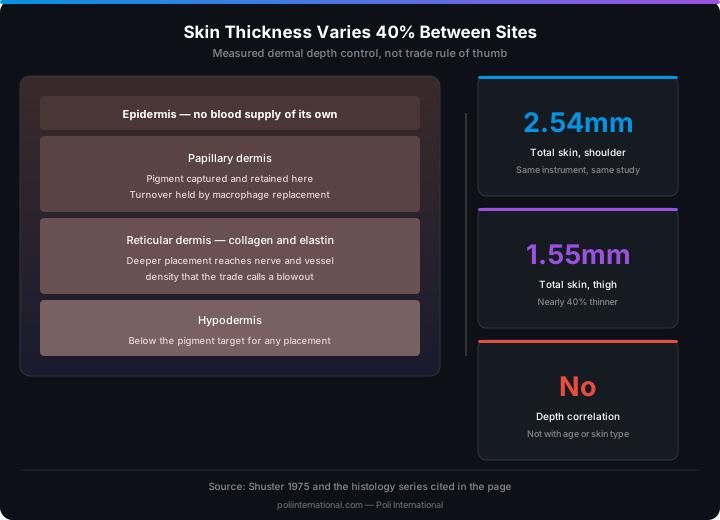
Artists are taught technique on skin as though it were one material. It is not. Between the shoulder and the thigh, measured on the same instrument in the same study, total skin thickness drops from 2.54 mm to 1.55 mm, a difference of nearly 40% [3]. Within the epidermis alone the ordering reverses depending on which sublayer you measure: the forearm has the thickest stratum corneum of the three sites in the histology series and the thinnest cellular epidermis [2]. A practitioner working to a fixed depth is therefore working to a different fraction of the tissue at every placement, and the error is systematic rather than random. This page deals only with what has been measured. The numbers that circulate in the trade without a source, including the usual statement of correct pigment depth in millimetres, are marked as such and left open.
Three Layers, And What Was Actually Measured
The clinical anatomy is standard and well summarised for practitioners in the dermatology nursing literature: epidermis, a stratified squamous epithelium with no blood supply of its own; dermis, the collagen and elastin connective layer that carries the vasculature, nerve endings and appendages; and hypodermis, subcutaneous fat and connective tissue [1]. What matters for body art is that these three are not measured the same way and are frequently conflated in trade material. Histological measurement of biopsies gives the epidermis in micrometres: in 71 volunteers, stratum corneum came out at 18.3 um on the dorsal forearm, 11.0 um on the shoulder and 14.9 um on the buttock, with the cellular epidermis beneath it at 56.6 um, 70.3 um and 81.5 um respectively [2]. High-frequency ultrasound measures epidermis and dermis together and gives millimetres: 2.54 mm suprascapular, 2.02 mm deltoid, 1.91 mm waist, 1.55 mm thigh across 205 women and 137 men aged 18 to 70 in three ethnic groups [3]. Those are two different quantities and they are not interchangeable. Set side by side (the two studies measured different sites), the epidermis comes out at only a few per cent of total skin thickness. Anyone quoting a single figure for 'skin thickness' without saying which measurement they mean is quoting something that could differ by a factor of twenty or more.
Two Findings That Contradict What The Trade Teaches
The histology series produced two results that cut directly against common studio assumptions. First, epidermal thickness was not correlated with age, and it was not correlated with skin type either [2]. The habit of adjusting technique because a client is older, or because they are darker or lighter, is not supported by epidermal measurement. What did correlate was body site, which explains most of the variation, along with a positive association between stratum corneum thickness and pigmentation and a negative association with years of smoking. Second, cellular epidermal thickness was significantly greater in males than in females and correlated positively with blood content [2]. That is a real difference and it is the opposite kind of variable from the one artists usually adjust for. The ultrasound study reinforces the point from the other direction: the measured thicknesses meant that a 1.5 mm needle inserted perpendicular to the surface would reach the dermis irrespective of sex, age, ethnicity and BMI [3]. Site dominates. Demographics, at these depths, largely do not.
Where Pigment Ends Up, And In Which Cells
The standard teaching is that ink must be placed in the dermis because the epidermis turns over and sheds anything left in it. In vivo two-photon fluorescence lifetime imaging says the reality is more complicated. Carbon black pigment was found intracellularly in both layers: in the epidermis within keratinocytes, dendritic cells and basal cells, and in the dermis within macrophages, mast cells and fibroblasts [6]. In this small series (one fresh tattoo and ten older than three years) it was present in the epidermis of 75% of the tattoos imaged, including tattoos applied up to nine years earlier, in skin showing no inflammation. In other words the epidermis of a long-healed tattoo is not pigment-free despite renewing continuously. The same study found carbon black still visible around the incision 84 days after a fresh application, which is a far longer epidermal recovery than the visible peel suggests. Independent atomic force microscopy of cryosectioned tattooed skin shows the same material distributed through the dermal collagen fibril network, and confirms that tattoo inks contain particles of sub-100 nm dimensions [8]. That paper also noted that manufacturers were not obliged to disclose the exact composition; in the EU this changed in January 2022, when REACH began requiring an ingredient list on the label. One further observation from the in vivo work has direct bearing on how tattooed skin behaves under a later piercing or a second pass: collagen I in tattooed skin showed higher directionality resembling scar tissue, with greater firmness and decreased elasticity [6].
Why The Tattoo Stays: Macrophage Turnover, Not Cell Longevity
The mechanism that keeps a tattoo in place was worked out in a mouse model with inducible macrophage ablation, and it is not the one most practitioners were taught. The skin myeloid cells that capture and retain tattoo pigment are exclusively dermal macrophages. When those macrophages are deleted, the pigment particles are released and recaptured by incoming macrophages, and the tattoo does not vanish. The particles can undergo successive cycles of capture, release and recapture without loss of the image, so long-term persistence relies on macrophage renewal rather than on any individual cell living for decades [7]. This is a mouse study and should be cited as one, but it explains two things a studio sees constantly: why a tattoo survives an inflammatory episode that destroys a great deal of local tissue, and why laser removal requires repeated sessions rather than one. Fragmenting the particles does not remove them; it only changes which cells can carry them and where they can be carried to.
? Open question, not established
What is the correct pigment depth in millimetres?
Almost every training source states a target depth, usually somewhere between one and two millimetres, and calls anything deeper a blowout. The anatomical premise is sound: pigment placed only in the epidermis will be carried off by turnover, and pigment placed into the hypodermis spreads laterally along a plane it cannot be held in. What is missing is a measurement. The published skin dimensions that exist are total thickness by site [3] and epidermal thickness in micrometres [2], and the pigment localisation work reports which cells hold pigment rather than at what depth in millimetres it was deposited [6] [8]. No source could be found that measured deposited pigment depth against outcome.
Held by: Needle and machine manufacturers in their training material, and practitioner consensus passed studio to studio. The specific millimetre figures vary between sources, which is itself a sign that none of them is derived.
What would settle it: A histological series measuring deposited pigment depth against retention and blowout at matched machine settings and body sites. Until one exists, the usable working rule is relative rather than absolute: the same hand depth is a different fraction of the tissue on a shoulder at 2.54 mm than on a thigh at 1.55 mm, and that ratio is measured even though the target is not.
Patrick's Note
"Fitzpatrick types I to VI appear in body art paperwork as a general skin classifier. They were not built for that. Fitzpatrick's own 1988 account explains that the scale was devised in 1975 to categorise lighter-skinned individuals for oral PUVA phototherapy dosing, after clinicians observed severe phototoxic reactions in patients whose tolerance had been assumed from phenotype, and he states directly that tolerance to oral PUVA could not be based solely on hair and eye colour. The original scale had four types; V and VI were added later [5]. The scale therefore measures sun reactivity, which is a genuine and useful input for laser work and for advising on sun exposure over a healing piece. It is not a measure of dermal thickness, of collagen density, of healing speed or of scar tendency, and the epidermal measurement series found no correlation between epidermal thickness and skin type at all [2]. Recording a Fitzpatrick type in a consultation is good practice for the things it predicts. Using it to decide needle depth is using a photobiology instrument as a ruler."
Founder & Piercing Expert
Poli International
Ageing Skin: What The Data Supports And What It Does Not
The reference measurement here is Shuster's 1975 series on forearm skin, which found that skin collagen decreases with age, that it is lower in females at every age, and that there is a direct relationship between skin collagen content and dermal thickness [4]. That is the basis for the real clinical observation that older skin offers a thinner, less resilient dermis. Two cautions follow directly from the same paper. First, the authors themselves note that variations in collagen density in disease limit the use of dermal thickness as a guide to collagen content, so the two are related but not substitutable. Second, the widely repeated figure of about one per cent collagen loss per year after the age of twenty is not what this study reports, and no source establishing that rate could be found; it should not be quoted as fact in client-facing material. Set against this, the epidermal measurement series found epidermal thickness uncorrelated with age [2]. The honest summary is that the dermis thins with age and the epidermis, as measured, does not, which is a more specific and more useful statement than 'skin gets thinner'.
Pre-Procedure Skin Assessment
A ten-step assessment carried out before the stencil goes on or the clamp closes. It is a structured look at the substrate, recorded in the client file so that a later outcome can be explained rather than guessed at.
- 1Identify the body site and treat site as the primary variable: total skin thickness at the suprascapular site is around 60% greater than at the thigh in the measured series [3]
- 2Inspect for existing tattooed skin in or adjacent to the working area, and expect reduced elasticity and scar-like collagen directionality there [6]
- 3Palpate for scar tissue, striae and previous surgical sites, where the dermal architecture is altered and pigment uptake becomes unpredictable
- 4Assess hydration and surface condition by sight and touch; a cracked or actively dermatitic surface is a reason to postpone, not a reason to work carefully
- 5Assess laxity by pinch, especially in older clients, where the dermis is thinner because collagen content has fallen [4]
- 6Record a Fitzpatrick type for what it actually predicts, sun reactivity and laser response, and not as a proxy for thickness or healing speed [5] [2]
- 7Note sebaceous and follicular density at the site; scalp, face and upper back behave differently from the forearm regardless of measured thickness
- 8Check for sun damage and current sunburn at the site; a compromised surface is a poor substrate and a healing site under UV is a separate problem
- 9For piercings, identify whether the intended channel passes through cartilage, and record it, because that determines the vascular supply available to the healing tissue rather than the dermal thickness
- 10Document the findings and any adjustment made, with the site named. Without a record there is no way later to distinguish a technique problem from a substrate problem

Substrate Errors And Where They Come From
Failure modes that trace back to a wrong model of the tissue rather than to a wrong hand.
- Working to one fixed depth across body sites — the same absolute depth is a different fraction of the tissue at 2.54 mm suprascapular and 1.55 mm thigh [3]
- Adjusting technique for the client's age on the assumption that the epidermis has thinned. Measured epidermal thickness showed no correlation with age [2]
- Adjusting for skin type on the same assumption. Epidermal thickness showed no correlation with skin type either [2]
- Quoting a one per cent per year collagen loss figure to clients. The 1975 study establishes that collagen falls with age, not that rate [4]
- Using Fitzpatrick type to set needle depth. It is a phototherapy dosing scale, devised for PUVA, not a thickness measure [5]
- Assuming the epidermis is pigment-free once a tattoo is healed. Pigment was found in the epidermis of 75% of tattoos up to nine years old [6]
- Treating a fresh tattoo as fully recovered when the surface stops peeling. Carbon black was still visible around the incision at 84 days [6]
- Tattooing over an existing tattoo without expecting altered mechanics — the collagen there has scar-like directionality, greater firmness and lower elasticity [6]
- Over-stretching the skin to work on it, which changes the geometry of the tissue under the needle and therefore the depth actually reached
- Treating cartilage as thin skin. The constraint at a cartilage site is vascular supply to the healing channel, not the millimetres of dermis over it
- Reading a single published 'skin thickness' figure without checking whether it is a histological epidermal measurement in micrometres or an ultrasound total in millimetres [2] [3]
Where Anatomy Sits In The Rules
No jurisdiction reviewed here sets an anatomy competency standard for body art practitioners. What exists is hygiene regulation that assumes anatomical understanding without specifying or testing it. This is a genuine gap, and it is reported as one rather than dressed up.
- EN 17169:2020 Tattooing, safe and hygienic practice is the European standard for the trade and sets hygiene requirements before, during and after tattooing, together with guidelines for practitioner conduct [10]
- That standard is built around infection control and client protection. It is not an anatomy or technique curriculum, and no European standard specifies dermal anatomy competency
- Practitioner licensing in most member states and in the UK sits with local authorities, whose criteria centre on premises and hygiene rather than on clinical knowledge
- In England, registration with the local authority under the Local Government (Miscellaneous Provisions) Act 1982 and its byelaws covers the practitioner and the premises; nothing in that process assesses understanding of skin structure. Scotland and Wales run their own licensing schemes
- The practical consequence: an artist can hold every licence the law requires and never have been examined on the tissue they work in
- Bloodborne pathogen training is the near-universal federal-facing requirement, and it treats intact skin as a barrier without teaching its structure
- Body art licensing is state by state, and the anatomy content of any required course varies from substantial to absent
- No federal standard establishes anatomical competency for tattooing or piercing
- Where states do require training hours, the hours are usually specified without a syllabus, so the content depends on the provider
- Client consent and record-keeping requirements are common; a documented pre-procedure skin assessment is rarely among them
- NSW Health's body art guidance places the obligation on the practitioner directly: they should only carry out procedures they have had appropriate training for and that are within their skills to complete [11]
- The same guidance requires the business to be registered with the local council and to meet planning and building standards before opening [11]
- NSW rules are prescriptive about sterilisation, sharps and skin antisepsis, and silent on anatomical knowledge, which is the same pattern as the EU and the US [11]
- Australian requirements are set state by state, so the training expectation differs across NSW, Victoria and Queensland
- Across the region the enforceable standards are infection control standards; anatomical competence is assumed rather than assessed
Patrick's Note
"The thing that surprised me most when I put this page together was how little of what the trade repeats about skin has ever been measured. I have been in this industry for a long time and I could have told you the shoulder is thicker than the thigh, but I could not have told you it is 2.54 millimetres against 1.55, because nobody in the trade quotes that number. They quote the depth figure instead, and that is the one with no study behind it. I would rather publish the gap than fill it with something that sounds authoritative. Where this matters to me practically is tension. A thin, mobile, lax site is where a rigid bar levers the channel every time the client moves or sleeps on it, and where a surface piercing walks out over months. That is a mechanical argument about the jewellery in the channel, not about the needle that made it, and it is the reason I created push-fit in the first place, because losing a jewel is better than tearing a piercing. Assess the substrate before you choose the geometry, write down what you found, and when a client asks how deep you work, tell them the honest answer: relative to the site, and by feel, because nobody has published the number. The client-facing version of this material is in the Journal at `/blog/skin-anatomy-body-art/`, and the Tattoo Needle Selector at `/needle-selector/` covers the configuration side."
Founder & Piercing Expert
Poli International
**Related Topics**
- »Anatomical Geometry, `/wiki/anatomical-geometry/`
- »Wound Healing Biology, `/wiki/wound-healing-biology/`
- »Needle Geometry Physics, `/wiki/needle-geometry-physics/`
- »Aftercare Formulation Chemistry, `/wiki/aftercare-formulation-chemistry/`
- »Piercing Migration and Rejection, `/wiki/piercing-migration-rejection/`
- »Skin Anatomy for Body Art (Journal), `/blog/skin-anatomy-body-art/`
- »Clinical Physics (Journal), `/blog/?category=Clinical%20Physics`
Common Questions
▸Why is understanding precise histological and ultrasound data critical for a body art professional working on different areas of the human body?
▸What specific anatomical measurements define skin thickness across different sites and what needle depth is required to reach the dermal layer?
▸How should a practitioner perform a pre-procedure assessment and which factors are most important when evaluating the tissue of a specific body site?
▸Why is it considered a technical error to adjust needle depth based on a client's age or skin type, and how should depth be determined?
▸What are the common misconceptions regarding healing timelines and pigment residence in the epidermis, and how can practitioners avoid these substrate errors?
▸What are the current regulatory requirements regarding anatomical knowledge for practitioners, and how do standards like EN 17169:2020 address clinical competency?
Anatomical Plates
Free Body Art Wall Posters
Print-ready reference posters for your studio or your own wall. Free to download and display, embed on your own site with attribution. Click any poster to preview it full size before downloading.
Page map · Skin Histology and Dermal Depth Control
- Skin Histology and Dermal Depth Control
- Critical Numbers
- Stratum corneum thickness
- Cellular epidermis over the same three sites
- Three Layers, And What Was Actually Measured
- Two Findings That Contradict What The Trade Teaches
- Where Pigment Ends Up, And In Which Cells
- Why The Tattoo Stays
- What is the correct pigment depth in millimetres?
- Fitzpatrick Typing Is A Phototherapy Dosing Tool
- Ageing Skin
- Pre-Procedure Skin Assessment
- Identify the body site and treat site as the primary variable
- Inspect for existing tattooed skin in or adjacent to the working area
- Substrate Errors And Where They Come From
- Working to one fixed depth across body sites
- Adjusting technique for the client's age on the assumption that the epidermis…
- Where Anatomy Sits In The Rules
- EU / UK
- United States
- Patrick's Note
Built from this page's own sections, so it always matches what is below. It is a map of the page, not a set of claims, and carries no citations of its own.
Technical Specifications
| Parameter | Standard / Value |
|---|---|
| Stratum corneum, dorsal forearm | 18.3 um (SD 4.9), histological measurement [2] |
| Stratum corneum, shoulder | 11.0 um (SD 2.2) [2] |
| Stratum corneum, buttock | 14.9 um (SD 3.4) [2] |
| Cellular epidermis, forearm / shoulder / buttock | 56.6 / 70.3 / 81.5 um (SD 11.5 / 13.6 / 15.7) [2] |
| Total skin thickness, suprascapular | 2.54 mm mean, 20 MHz ultrasound, n=342 [3] |
| Total skin thickness, deltoid | 2.02 mm mean [3] |
| Total skin thickness, waist | 1.91 mm mean [3] |
| Total skin thickness, thigh | 1.55 mm mean [3] |
| Needle length that reaches the dermis (authors' conclusion) | 1.5 mm perpendicular, regardless of sex, age, ethnicity and BMI [3] |
| Epidermal thickness vs age | No correlation found [2] |
| Epidermal thickness vs skin type | No correlation found [2] |
| Sex difference in cellular epidermis | Significantly greater in males than females [2] |
| Dermal collagen and age | Forearm skin collagen decreases with age and is lower in females at all ages [4] |
| Tattoo pigment localisation | Exclusively intracellular: keratinocytes, dendritic and basal cells (epidermis); macrophages, mast cells, fibroblasts (dermis) [6] |
| Pigment persistence in epidermis | Present in 75% of tattoos up to 9 years old, in non-inflamed skin (small series: 1 fresh, 10 older tattoos) [6] |
| Tattooed dermis mechanics | Collagen I directionality resembling scar tissue; greater firmness, decreased elasticity [6] |
| Pigment retention mechanism | Dermal macrophage capture-release-recapture cycles; persistence via macrophage renewal (mouse model) [7] |
| Tattoo ink particle size | Sub-100 nm particles present [8]; EU ingredient labelling required since January 2022 |
| Fitzpatrick scale origin | Devised 1975 (types I to IV) for oral PUVA dosing, V and VI added later; a sun-reactivity scale, not a thickness measure [5] |
Revision Deck
19 cards, taken directly from this page's specifications. Nothing here is generated: the deck is the same reviewed data as the table above. See the full table.
Printable wall chart (A4) — the critical numbers, the do-nots and the applicable standards on one page for the bench.
References
- [1]Kolarsick PAJ, Kolarsick MA, Goodwin C. Anatomy and Physiology of the Skin. J Dermatol Nurses Assoc. 2011;3(4):203-213.https://doi.org/10.1097/JDN.0b013e3182274a98
- [2]Sandby-Møller J, Poulsen T, Wulf HC. Epidermal thickness at different body sites: relationship to age, gender, pigmentation, blood content, skin type and smoking habits. Acta Derm Venereol. 2003;83(6):410-3.https://doi.org/10.1080/00015550310015419
- [3]Laurent A, Mistretta F, Bottigioli D, Dahel K, Goujon C, Nicolas JF, Hennino A, Laurent PE. Echographic measurement of skin thickness in adults by high frequency ultrasound to assess the appropriate microneedle length for intradermal delivery of vaccines. Vaccine. 2007;25(34):6423-30.https://doi.org/10.1016/j.vaccine.2007.05.046
- [4]Shuster S, Black MM, McVitie E. The influence of age and sex on skin thickness, skin collagen and density. Br J Dermatol. 1975;93(6):639-43.https://doi.org/10.1111/j.1365-2133.1975.tb05113.x
- [5]Fitzpatrick TB. The Validity and Practicality of Sun-Reactive Skin Types I Through VI. Arch Dermatol. 1988;124(6):869-871.https://doi.org/10.1001/archderm.1988.01670060015008
- [6]Kröger M, Schleusener J, Lademann J, Meinke MC, Jung S, Darvin ME. Tattoo Pigments Are Localized Intracellularly in the Epidermis and Dermis of Fresh and Old Tattoos: In vivo Study Using Two-Photon Excited Fluorescence Lifetime Imaging. Dermatology. 2023;239(3):478-493.https://doi.org/10.1159/000529577
- [7]Baranska A, Shawket A, Jouve M, et al. Unveiling skin macrophage dynamics explains both tattoo persistence and strenuous removal. J Exp Med. 2018;215(4):1115-1133.https://doi.org/10.1084/jem.20171608
- [8]Grant CA, Twigg PC, Baker R, Tobin DJ. Tattoo ink nanoparticles in skin tissue and fibroblasts. Beilstein J Nanotechnol. 2015;6:1183-91.https://doi.org/10.3762/bjnano.6.120
- [9]Winter GD. Formation of the scab and the rate of epithelization of superficial wounds in the skin of the young domestic pig. Nature. 1962;193:293-4.https://doi.org/10.1038/193293a0
- [10]EN 17169:2020. Tattooing, Safe and hygienic practice. European Committee for Standardization.https://standards.iteh.ai/catalog/standards/cen/275ed7ba-d629-4594-b34b-2adc90db4a86/en-17169-2020
- [11]NSW Health. Body art and tattooing businesses. Skin penetration infection control practices, November 2025.https://www.health.nsw.gov.au/environment/factsheets/Factsheets/tattooing.pdf
- [12]Junker JP, Kamel RA, Caterson EJ, Eriksson E. Clinical Impact Upon Wound Healing and Inflammation in Moist, Wet, and Dry Environments. Adv Wound Care (New Rochelle). 2013;2(7):348-356.https://doi.org/10.1089/wound.2012.0412
Put Science Into Practice
This technical standard is the architectural foundation for our professional analytical tools.