Skin Is Not a Static Canvas. Here Is What Happens to Body Art Over Decades.
Key Takeaways:
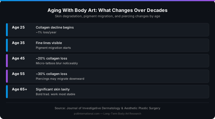
» Dermal collagen declines at roughly 1% per year after age 25. By age 50, the skin has 25 to 30% less structural protein than when the tattoo was first applied.
» Pigment migrates over decades through the macrophage relay cycle. Black ink is the most stable. Yellow, orange, and pastel pigments migrate and fade faster. Fine-line micro tattoos blur more visibly than bold traditional work.
» Piercings on aging tissue face higher risk of migration and rejection as the dermal scaffold thins. Lighter jewellery materials such as titanium and BioFlex reduce the mechanical load on the fistula.
» Sun protection is the single most effective intervention for tattoo longevity. Daily SPF 30+ sunscreen reduces UV-A penetration by roughly 95% and slows both pigment fading and collagen breakdown.
» New body art on older skin is safe with adjusted technique: shallower needle depth, extended healing timelines, and medication interaction screening.
1. What Happens to Skin as It Ages
Skin is a living organ that remodels continuously, and the remodelling rate changes with age in predictable ways. After roughly age 25, dermal collagen declines at approximately 1% per year. By age 50, the dermis has lost roughly 25 to 30% of its collagen content compared to young adult skin. Collagen provides the structural scaffold that holds tattoo pigment in place and gives pierced tissue its resistance to tearing and migration.
Elastin fibres, which give skin its ability to stretch and recoil, degrade significantly with photoaging. Sun-exposed areas show far more elastin degradation than protected skin. When elastin breaks down, the skin sags and does not return to its original position after being stretched or loaded with jewellery weight. The epidermis thins by roughly 6 to 8% per decade, and the dermal-epidermal junction flattens, making the skin more fragile. A piercing that sat comfortably in dense dermal tissue at age 25 may sit in thinner, less supportive tissue at age 55.
Dermal capillary density also decreases with age. The result is slower wound healing, reduced immune surveillance, and a longer inflammatory phase after any skin injury. A tattoo that healed in four weeks at age 25 may take six to eight weeks at age 60.
2. Tattoos Over Decades: Pigment Migration, Fading, and Distortion
Pigment particles do not stay exactly where the needle placed them. Over decades, macrophages containing pigment die, release their contents, and are replaced by new macrophages that pick up the pigment in slightly different positions. This macrophage relay cycle, combined with the slow degradation of the collagen matrix, causes pigment to spread laterally, blurring sharp lines and reducing edge definition.
The rate of migration varies by ink colour. Black carbon-based inks are the most stable. Yellow, orange, and pastel pigments migrate faster because they absorb more UV energy and the pigment particles are typically smaller and more mobile in the dermal matrix. Blue and green phthalocyanine pigments tend to outlast reds, yellows, and oranges because the particle sizes are larger and less UV energy is transferred to the pigment molecule. A black-and-blue traditional tattoo will usually retain legibility longer than a watercolour piece using predominantly warm tones.
Fine-line and micro tattoos are particularly vulnerable. These styles rely on precise pigment placement with minimal ink volume. When the pigment migrates even one or two macrophage-diameter distances, the fine detail blurs. A micro tattoo that looked sharp at year two may be an unreadable smudge at year twenty. This is not a failure of the artist: it is the biology of the dermis.
When skin sags, the tattoo distorts. A geometric mandala on a flat twenty-year-old ribcage may become an oval shape on a sixty-year-old torso. There is no fix for this: it is gravity acting on tissue that has lost its structural proteins.
3. Piercings and Aging: Migration, the Cheese-Cutter Effect, and When to Retire
Piercings age differently than tattoos because they involve a fistula, a healed channel of scar tissue that forms around the jewellery. That channel exists in dynamic equilibrium with the surrounding tissue. When the surrounding tissue thins, the fistula changes.
As dermal collagen declines, the tissue supporting a piercing becomes less dense. Jewellery that sat comfortably for twenty years may begin to migrate downward, a slow version of the cheese-cutter effect where the jewellery post gradually cuts through thinning tissue. This is most common in surface piercings and ear cartilage piercings with heavy jewellery. Earlobes lose collagen and elastin faster than most other body sites; heavy earrings worn over decades can elongate the piercing channel from a round hole to a permanent slit.
Retiring a piercing is a clinical decision, not a failure. Signs that a piercing should be retired include visible thinning of the tissue bridge above the jewellery to less than 2 mm, migration exceeding 3 mm from the original placement, and recurrent infections in the same fistula. For aging clients considering retirement, soft-tissue fistulas usually close within days to weeks. Cartilage piercings may leave a permanent channel. Earlobe slits typically require surgical repair.